Explore
 Validate
 Learn
Learn Western blot
 Immunocytochemistry
Antibody data
- Antibody Data
- Antigen structure
- References [2]
- Comments [0]
- Validations
- Immunocytochemistry [1]
- Immunohistochemistry [2]
- Other assay [1]
Submit
Validation data
Reference
Comment
Report error
- Product number
- PA5-27607 - Provider product page

- Provider
- Invitrogen Antibodies
- Product name
- DIAPH1 Polyclonal Antibody
- Antibody type
- Polyclonal
- Antigen
- Recombinant full-length protein
- Description
- Recommended positive controls: H1299, Raji. Predicted reactivity: Mouse (93%), Rat (94%), Pig (88%). Store product as a concentrated solution. Centrifuge briefly prior to opening the vial.
- Reactivity
- Human, Mouse
- Host
- Rabbit
- Isotype
- IgG
- Vial size
- 100 μL
- Concentration
- 1 mg/mL
- Storage
- Store at 4°C short term. For long term storage, store at -20°C, avoiding freeze/thaw cycles.
Submitted references Formin Activity and mDia1 Contribute to Maintain Axon Initial Segment Composition and Structure.
Extension of the clinical and molecular phenotype of DIAPH1-associated autosomal dominant hearing loss (DFNA1).
Zhang W, Ciorraga M, Mendez P, Retana D, Boumedine-Guignon N, Achón B, Russier M, Debanne D, Garrido JJ
Molecular neurobiology 2021 Dec;58(12):6153-6169
Molecular neurobiology 2021 Dec;58(12):6153-6169
Extension of the clinical and molecular phenotype of DIAPH1-associated autosomal dominant hearing loss (DFNA1).
Neuhaus C, Lang-Roth R, Zimmermann U, Heller R, Eisenberger T, Weikert M, Markus S, Knipper M, Bolz HJ
Clinical genetics 2017 Jun;91(6):892-901
Clinical genetics 2017 Jun;91(6):892-901
No comments: Submit comment
Supportive validation
- Submitted by
- Invitrogen Antibodies (provider)
- Main image
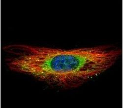
- Experimental details
- Immunofluorescent analysis of DIAPH1 in methanol-fixed HeLa cells using a DIAPH1 polyclonal antibody (Product # PA5-27607) (Green) at a 1:500 dilution. Alpha-tubulin filaments were labeled with Product # PA5-29281 (Red) at a 1:2000.

Supportive validation
- Submitted by
- Invitrogen Antibodies (provider)
- Main image
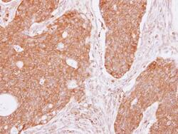
- Experimental details
- DIAPH1 Polyclonal Antibody detects DIAPH1 protein at cytoplasm on human colon carcinoma by immunohistochemical analysis. Sample: Paraffin-embedded colon carcinoma. DIAPH1 Polyclonal Antibody (Product # PA5-27607) dilution: 1:250. Antigen Retrieval: EDTA based buffer, pH 8.0, 15 min.

- Submitted by
- Invitrogen Antibodies (provider)
- Main image
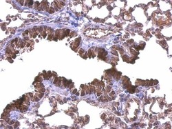
- Experimental details
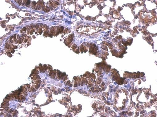
- DIAPH1 Polyclonal Antibody detects DIAPH1 protein at membrane on mouse lung by immunohistochemical analysis. Sample: Paraffin-embedded mouse lung. DIAPH1 Polyclonal Antibody (Product # PA5-27607) dilution: 1:500. Antigen Retrieval: EDTA based buffer, pH 8.0, 15 min.
Supportive validation
- Submitted by
- Invitrogen Antibodies (provider)
- Main image
- Experimental details
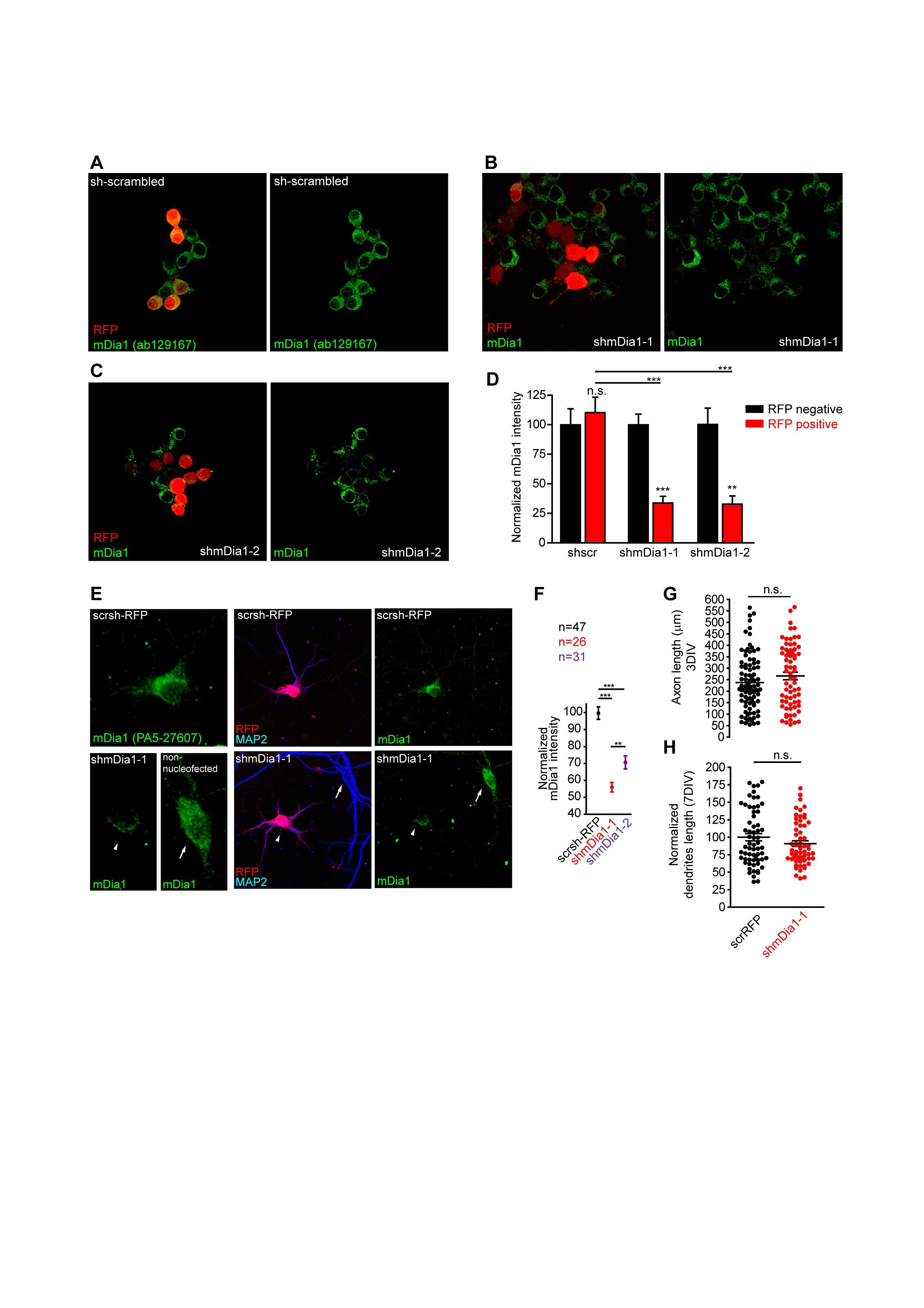
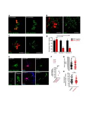
- Supplementary Figure 5. ( A-C ) Neuro2a cells transfected with srambled (A), shmDia1-1 (B) or shmDia1-2 (C) interference shRNAs for 3 days. Transfected cells are identified by RFP signal (red) and mDia1 knockout validated antibody (ab129167, Abcam) signal is show in green. ( D ) Quantification of mDia1 signal in transfected (red bars) or non-transfected (black bars) Neuro2a cells (n=50 cells/experimental condition). Data are represented as the mean +- SEM. n.s., not significant, ** p < 0.01***, p < 0.0001, Mann-Whitney test. ( E ) 10 DIV hippocampal neurons nucleofected with scrambled interference RNA (scrsh-RFP) or mDia1 interference RNAs (shmDia1-1, shmDia1-2). Nucleofected neurons were identified based on RFP fluorescence (magenta). Neurons were stained with mDia1 (green) and MAP2 (blue) antibodies. Scale bar = 20 mum. ( F ) mDia1 fluorescence intensity was quantified in the soma of neurons nucleofected with scrsh-RFP, shmDia1-1 or shmDia1-2 plasmids. Inserts in D show magnifications of mDia1 staining in soma. **p < 0.01, ***p < 0.001, One-way analysis of variance, Tukey's multiple comparison test. Data in graphs were acquired from three independent experiments and represented as the mean +- SEM. ( G, H ) Axonal and dendritic growth is not affected by mDia1 interference RNA. Hippocampal neurons were nucleofected before plating with shmDia1-1 interference RNA and kept for 3 DIV (G) to analyze axonal length using the tau1 axonal marker, or 7 DIV to analyze total dendritic le