Explore
 Validate
 Learn
Learn Western blot
 ELISA
ELISA Immunocytochemistry
Antibody data
- Antibody Data
- Antigen structure
- References [0]
- Comments [0]
- Validations
- Western blot [2]
- Immunohistochemistry [1]
- Flow cytometry [1]
Submit
Validation data
Reference
Comment
Report error
- Product number
- AP51968PU-N - Provider product page

- Provider
- Acris Antibodies GmbH
- Proper citation
- Acris Antibodies GmbH Cat#AP51968PU-N, RRID:AB_11150650
- Product name
- anti GSTP1 / GST3 (Center)
- Antibody type
- Polyclonal
- Antigen
- KLH conjugated synthetic peptide between 104~134 amino acids from the Center region of Human GSTP1 / GST3.
- Reactivity
- Human
- Host
- Rabbit
- Vial size
- 0.1 mg
- Concentration
- 0.25 mg/ml
No comments: Submit comment
Supportive validation
- Submitted by
- Acris Antibodies GmbH (provider)
- Main image
- Experimental details
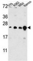
- Western blot analysis of GSTP1 / GST3 Antibody (Center) Cat.-No AP51968PU-N in Y79, K562, WIDr, Ramos cell line lysates (35ug/lane). GSTP1 (arrow) was detected using the purified Pab.

- Submitted by
- Acris Antibodies GmbH (provider)
- Main image

- Experimental details
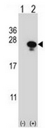
- Western blot analysis of GSTP1 (arrow) using GSTP1 / GST3 Antibody (Center) Cat.-No AP51968PU-N. 293 cell lysates (2 ug/lane) either nontransfected (Lane 1) or transiently transfected (Lane 2) with the GSTP1 gene.
Supportive validation
- Submitted by
- Acris Antibodies GmbH (provider)
- Main image
- Experimental details
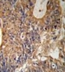
- Immunohistochemistry analysis in formalin fixed and paraffin embedded human colon carcinoma reacted with GSTP1 / GST3 Antibody (Center) Cat.-No AP51968PU-N followed by peroxidase conjugation of the secondary antibody and DAB staining.

Supportive validation
- Submitted by
- Acris Antibodies GmbH (provider)
- Main image

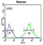
- Experimental details
- Flow cytometric analysis of Ramos cells using GSTP1 / GST3 Antibody (Center) Cat.-No AP51968PU-N (right histogram) compared to a negative control cell (left histogram). FITC-conjugated goat-anti-rabbit secondary antibodies were used for the analysis.