Explore
 Validate
 Learn
Learn Western blot
Antibody data
- Antibody Data
- Antigen structure
- References [1]
- Comments [0]
- Validations
- Western blot [2]
- Immunocytochemistry [2]
Submit
Validation data
Reference
Comment
Report error
- Product number
- MAB6455 - Provider product page

- Provider
- R&D Systems
- Product name
- Human Glutathione S-Transferase pi 1/GSTP1 Antibody
- Antibody type
- Monoclonal
- Description
- Protein A or G purified from hybridoma culture supernatant. Detects human Glutathione S-Transferase pi 1/GSTP1 in direct ELISAs and Western blots. Detects mouse and rat Glutathione S-Transferase pi 1/GSTP1 in Western Blots.
- Reactivity
- Human
- Host
- Mouse
- Conjugate
- Unconjugated
- Antigen sequence
AAC51280- Isotype
- IgG
- Antibody clone number
- 800027
- Vial size
- 100 ug
- Concentration
- LYOPH
- Storage
- Use a manual defrost freezer and avoid repeated freeze-thaw cycles. 12 months from date of receipt, -20 to -70 °C as supplied. 1 month, 2 to 8 °C under sterile conditions after reconstitution. 6 months, -20 to -70 °C under sterile conditions after reconstitution.
Submitted references Mining the nucleus accumbens proteome for novel targets of alcohol self-administration in male C57BL/6J mice.
Faccidomo S, Swaim KS, Saunders BL, Santanam TS, Taylor SM, Kim M, Reid GT, Eastman VR, Hodge CW
Psychopharmacology 2018 Jun;235(6):1681-1696
Psychopharmacology 2018 Jun;235(6):1681-1696
No comments: Submit comment
Supportive validation
- Submitted by
- R&D Systems (provider)
- Main image
- Experimental details
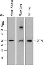
- Detection of Human, Mouse, and Rat Glutathione S-Transferase pi 1/GSTP1 by Western Blot. Western blot shows lysates of human placenta tissue, mouse lung tissue, and rat lung tissue. PVDF membrane was probed with 0.25 µg/mL of Mouse Anti-Human Glutathione S-Transferase pi 1/GSTP1 Monoclonal Antibody (Catalog # MAB6455) followed by HRP-conjugated Anti-Mouse IgG Secondary Antibody (Catalog # HAF018). A specific band was detected for Glutathione S-Transferase pi 1/GSTP1 at approximately 25 kDa (as indicated). This experiment was conducted under reducing conditions and using Immunoblot Buffer Group 1.

- Submitted by
- R&D Systems (provider)
- Main image
- Experimental details
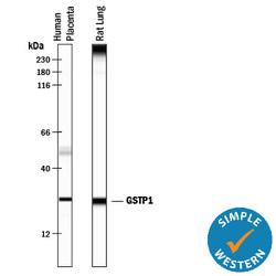
- Detection of Human and Rat Glutathione S-Transferase pi 1/GSTP1 by Simple WesternTM. Simple Western lane view shows lysates of human placenta tissue and rat brain tissue, loaded at 0.5 mg/mL. A specific band was detected for Glutathione S-Transferase pi 1/GSTP1 at approximately 26 kDa (as indicated) using 12.5 µg/mL of Mouse Anti-Human Glutathione S-Transferase pi 1/GSTP1 Monoclonal Antibody (Catalog # MAB6455). This experiment was conducted under reducing conditions and using the 12-230 kDa separation system.

Supportive validation
- Submitted by
- R&D Systems (provider)
- Main image
- Experimental details
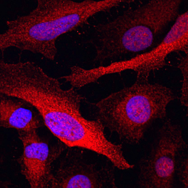
- Glutathione S-Transferase pi 1/GSTP1 in BG01V Human Embryonic Stem Cells. Glutathione S-Transferase pi 1/GSTP1 was detected in immersion fixed BG01V human embryonic stem cells differentiated into hepatocytes using Mouse Anti-Human Glutathione S-Transferase pi 1/GSTP1 Monoclonal Antibody (Catalog # MAB6455) at 10 µg/mL for 3 hours at room temperature. Cells were stained using the NorthernLights™ 557-conjugated Anti-Mouse IgG Secondary Antibody (red; Catalog # NL007) and counterstained with DAPI (blue). Specific staining was localized to cytoplasm. View our protocol for Fluorescent ICC Staining of Stem Cells on Coverslips.

- Submitted by
- R&D Systems (provider)
- Main image

- Experimental details
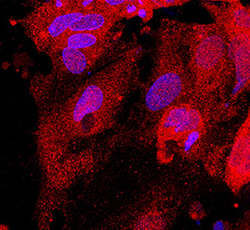
- Glutathione S-Transferase pi 1/GSTP1 in HeLa Human Cell Line. Glutathione S-Transferase pi 1/GSTP1 was detected in immersion fixed HeLa human cervical epithelial carcinoma cell line using Mouse Anti-Human Glutathione S-Transferase pi 1/GSTP1 Monoclonal Antibody (Catalog # MAB6455) at 10 µg/mL for 3 hours at room temperature. Cells were stained using the NorthernLights™ 557-conjugated Anti-Mouse IgG Secondary Antibody (red; Catalog # NL007) and counterstained with DAPI (blue). Specific staining was localized to cytoplasm. View our protocol for Fluorescent ICC Staining of Cells on Coverslips.