Explore
 Validate
 Learn
Learn Western blot
Antibody data
- Antibody Data
- Antigen structure
- References [0]
- Comments [0]
- Validations
- Western blot [2]
- Immunohistochemistry [1]
- Flow cytometry [1]
Submit
Validation data
Reference
Comment
Report error
- Product number
- ACR-050-200UL - Provider product page

- Provider
- Invitrogen Antibodies
- Product name
- CRF1/CRHR1 (extracellular) Polyclonal Antibody
- Antibody type
- Polyclonal
- Antigen
- Other
- Reactivity
- Human, Mouse, Rat
- Host
- Rabbit
- Isotype
- IgG
- Vial size
- 200 µL
- Concentration
- 0.85 mg/mL
- Storage
- -20° C, Avoid Freeze/Thaw Cycles
No comments: Submit comment
Supportive validation
- Submitted by
- Invitrogen Antibodies (provider)
- Main image

- Experimental details
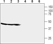
- Western blot analysis of mouse brain membrane (lanes 1 and 4), rat brain membrane (lane 2 and 5) and rat cortex lysate (lane 3 and 6): - 1-3. Anti-CRF1/CRHR1 (extracellular) Antibody (#ACR-050), (1:200).4-6. Anti-CRF1/CRHR1 (extracellular) Antibody , preincubated with CRF1/CRHR1 (extracellular) Blocking Peptide (#BLP-CR050).
- Submitted by
- Invitrogen Antibodies (provider)
- Main image
- Experimental details
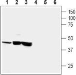
- Western blot analysis of human neuroblastoma (SH-SY5Y) (lanes 1 and 4), human brain astrocytoma (CCF-STTGI) (lanes 2 and 5) and rat pheochromocytoma (PC12) (lanes 3 and 6): - 1-3. Anti-CRF1/CRHR1 (extracellular) Antibody (#ACR-050), (1:200).4-6. Anti-CRF1/CRHR1 (extracellular) Antibody , preincubated with CRF1/CRHR1 (extracellular) Blocking Peptide (#BLP-CR050).

Supportive validation
- Submitted by
- Invitrogen Antibodies (provider)
- Main image
- Experimental details
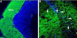
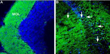
- Expression of Corticotropin-releasing factor receptor 1 in mouse brain - Immunohistochemical staining of mouse hippocampus and mouse cerebellum using Anti-CRF1/CRHR1 (extracellular) Antibody (#ACR-050), (1:600). A. CRF1 staining (green) in the cerebellum appears in cerebellar molecular layer (MOL). B. CRF1staining (green)in the hippocampus appears in soma (horizontal arrows) and dendrites of pyramidal neurons (arrow). In both panels DAPI was used as the counterstain.
Supportive validation
- Submitted by
- Invitrogen Antibodies (provider)
- Main image
- Experimental details
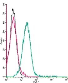
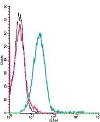
- Cell surface detection of Corticotropin-releasing factor receptor 1 by indirect flow cytometry in live intact mouse P815 mastocytoma cells: - (black line) cells. (red) Cells + goat- Anti-rabbit-FITC. (green) Cells + Anti-CRF1/CRHR1 (extracellular) Antibody (#ACR-050), (2.5μg) + goat- Anti-rabbit-FITC.