 Explore
 Validate
 Learn
Learn436000
antibody from Invitrogen Antibodies
Targeting: MMP2
CLG4, CLG4A, TBE-1
 Western blot
 Immunocytochemistry
 Immunoprecipitation Immunohistochemistry Flow cytometry Other assay
Antibody data
- Antibody Data
- Antigen structure
- References [11]
- Comments [0]
- Validations
- Immunocytochemistry [2]
- Immunohistochemistry [1]
- Flow cytometry [2]
- Other assay [10]
Submit
Validation data
Reference
Comment
Report error
- Product number
- 436000 - Provider product page

- Provider
- Invitrogen Antibodies
- Product name
- MMP2 Monoclonal Antibody (101)
- Antibody type
- Monoclonal
- Antigen
- Recombinant full-length protein
- Reactivity
- Human, Mouse, Rat, Rabbit
- Host
- Mouse
- Isotype
- IgG
- Antibody clone number
- 101
- Vial size
- 100 μg
- Concentration
- 0.5 mg/mL
- Storage
- -20°C
Submitted references EIF5A2 Is Involved in the Biological Process of Cervical Cancer Cells through AGR2.
Adiponectin and Leptin Exert Antagonizing Effects on HUVEC Tube Formation and Migration Modulating the Expression of CXCL1, VEGF, MMP-2 and MMP-9.
A hypoxia-induced Rab pathway regulates embryo implantation by controlled trafficking of secretory granules.
Expression of metalloproteinases MMP-2 and MMP-9 is associated to the presence of androgen receptor in epithelial ovarian tumors.
MMP-2 and MMP-9 as prognostic markers for the early detection of urinary bladder cancer.
Amphiregulin Regulates Phagocytosis-Induced Cell Death in Monocytes via EGFR and Matrix Metalloproteinases.
Lactobacillus casei Strain Shirota Enhances the In Vitro Antiproliferative Effect of Geniposide in Human Oral Squamous Carcinoma HSC-3 Cells.
The Activity of Matrix Metalloproteinases (MMP-2, MMP-9) and Their Tissue Inhibitors (TIMP-1, TIMP-3) in the Cerebral Cortex and Hippocampus in Experimental Acanthamoebiasis.
PARP-1 depletion in combination with carbon ion exposure significantly reduces MMPs activity and overall increases TIMPs expression in cultured HeLa cells.
Diurnal variation of tight junction integrity associates inversely with matrix metalloproteinase expression in Xenopus laevis corneal epithelium: implications for circadian regulation of homeostatic surface cell desquamation.
Lung dysfunction causes systemic hypoxia in estrogen receptor beta knockout (ERbeta-/-) mice.
Shen X, Li L, He Y, Lv X, Ma J
Pharmacology 2022;107(7-8):376-385
Pharmacology 2022;107(7-8):376-385
Adiponectin and Leptin Exert Antagonizing Effects on HUVEC Tube Formation and Migration Modulating the Expression of CXCL1, VEGF, MMP-2 and MMP-9.
Nigro E, Mallardo M, Polito R, Scialò F, Bianco A, Daniele A
International journal of molecular sciences 2021 Jul 13;22(14)
International journal of molecular sciences 2021 Jul 13;22(14)
A hypoxia-induced Rab pathway regulates embryo implantation by controlled trafficking of secretory granules.
Bhurke A, Kannan A, Neff A, Ma Q, Laws MJ, Taylor RN, Bagchi MK, Bagchi IC
Proceedings of the National Academy of Sciences of the United States of America 2020 Jun 23;117(25):14532-14542
Proceedings of the National Academy of Sciences of the United States of America 2020 Jun 23;117(25):14532-14542
Expression of metalloproteinases MMP-2 and MMP-9 is associated to the presence of androgen receptor in epithelial ovarian tumors.
Morales-Vásquez F, Castillo-Sánchez R, Gómora MJ, Almaraz MÁ, Pedernera E, Pérez-Montiel D, Rendón E, López-Basave HN, Román-Basaure E, Cuevas-Covarrubias S, Maldonado-Cubas J, Villa A, Mendez C
Journal of ovarian research 2020 Jul 28;13(1):86
Journal of ovarian research 2020 Jul 28;13(1):86
MMP-2 and MMP-9 as prognostic markers for the early detection of urinary bladder cancer.
Fouad H, Salem H, Ellakwa DE, Abdel-Hamid M
Journal of biochemical and molecular toxicology 2019 Apr;33(4):e22275
Journal of biochemical and molecular toxicology 2019 Apr;33(4):e22275
Amphiregulin Regulates Phagocytosis-Induced Cell Death in Monocytes via EGFR and Matrix Metalloproteinases.
Platen C, Dreschers S, Reiss LK, Wappler J, Orlikowsky TW
Mediators of inflammation 2018;2018:4310419
Mediators of inflammation 2018;2018:4310419
Lactobacillus casei Strain Shirota Enhances the In Vitro Antiproliferative Effect of Geniposide in Human Oral Squamous Carcinoma HSC-3 Cells.
Qian Y, Song JL, Sun P, Yi R, Liu H, Feng X, Park KY, Zhao X
Molecules (Basel, Switzerland) 2018 May 3;23(5)
Molecules (Basel, Switzerland) 2018 May 3;23(5)
The Activity of Matrix Metalloproteinases (MMP-2, MMP-9) and Their Tissue Inhibitors (TIMP-1, TIMP-3) in the Cerebral Cortex and Hippocampus in Experimental Acanthamoebiasis.
Łanocha-Arendarczyk N, Baranowska-Bosiacka I, Gutowska I, Kolasa-Wołosiuk A, Kot K, Łanocha A, Metryka E, Wiszniewska B, Chlubek D, Kosik-Bogacka D
International journal of molecular sciences 2018 Dec 19;19(12)
International journal of molecular sciences 2018 Dec 19;19(12)
PARP-1 depletion in combination with carbon ion exposure significantly reduces MMPs activity and overall increases TIMPs expression in cultured HeLa cells.
Ghorai A, Sarma A, Chowdhury P, Ghosh U
Radiation oncology (London, England) 2016 Sep 22;11(1):126
Radiation oncology (London, England) 2016 Sep 22;11(1):126
Diurnal variation of tight junction integrity associates inversely with matrix metalloproteinase expression in Xenopus laevis corneal epithelium: implications for circadian regulation of homeostatic surface cell desquamation.
Wiechmann AF, Ceresa BP, Howard EW
PloS one 2014;9(11):e113810
PloS one 2014;9(11):e113810
Lung dysfunction causes systemic hypoxia in estrogen receptor beta knockout (ERbeta-/-) mice.
Morani A, Barros RP, Imamov O, Hultenby K, Arner A, Warner M, Gustafsson JA
Proceedings of the National Academy of Sciences of the United States of America 2006 May 2;103(18):7165-9
Proceedings of the National Academy of Sciences of the United States of America 2006 May 2;103(18):7165-9
No comments: Submit comment
Supportive validation
- Submitted by
- Invitrogen Antibodies (provider)
- Main image

- Experimental details
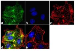
- Immunofluorescence analysis of MMP-2/CLG4A was performed using 70% confluent log phase MCF-7 cells. The cells were fixed with 4% paraformaldehyde for 10 minutes, permeabilized with 0.1% Triton™ X-100 for 10 minutes, and blocked with 1% BSA for 1 hour at room temperature. The cells were labeled with MMP-2/CLG4A (101) Mouse Monoclonal Antibody (Product # 436000) at 2 µg/mL in 0.1% BSA and incubated for 3 hours at room temperature and then labeled with Goat anti-Mouse IgG (H+L) Superclonal™ Secondary Antibody, Alexa Fluor® 488 conjugate (Product # A28175) at a dilution of 1:2000 for 45 minutes at room temperature (Panel a: green). Nuclei (Panel b: blue) were stained with SlowFade® Gold Antifade Mountant with DAPI (Product # S36938). F-actin (Panel c: red) was stained with Alexa Fluor® 555 Rhodamine Phalloidin (Product # R415, 1:300). Panel d represents the merged image showing cytoplasmic localization. Panel e shows the no primary antibody control. The images were captured at 60X magnification.

- Submitted by
- Invitrogen Antibodies (provider)
- Main image
- Experimental details
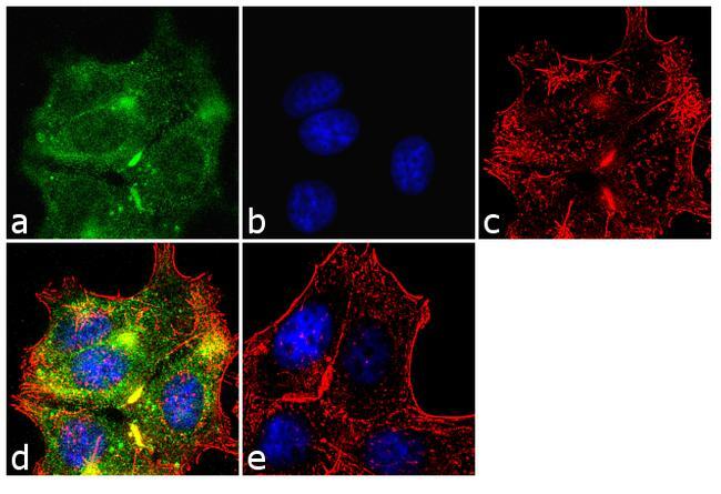
- Immunofluorescence analysis of MMP-2/CLG4A was performed using 70% confluent log phase MCF-7 cells. The cells were fixed with 4% paraformaldehyde for 10 minutes, permeabilized with 0.1% Triton™ X-100 for 10 minutes, and blocked with 1% BSA for 1 hour at room temperature. The cells were labeled with MMP-2/CLG4A (101) Mouse Monoclonal Antibody (Product # 436000) at 2 µg/mL in 0.1% BSA and incubated for 3 hours at room temperature and then labeled with Goat anti-Mouse IgG (H+L) Superclonal™ Secondary Antibody, Alexa Fluor® 488 conjugate (Product # A28175) at a dilution of 1:2000 for 45 minutes at room temperature (Panel a: green). Nuclei (Panel b: blue) were stained with SlowFade® Gold Antifade Mountant with DAPI (Product # S36938). F-actin (Panel c: red) was stained with Alexa Fluor® 555 Rhodamine Phalloidin (Product # R415, 1:300). Panel d represents the merged image showing cytoplasmic localization. Panel e shows the no primary antibody control. The images were captured at 60X magnification.
Supportive validation
- Submitted by
- Invitrogen Antibodies (provider)
- Main image
- Experimental details
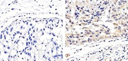
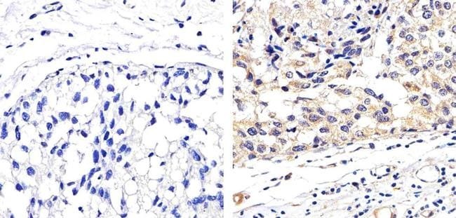
- Immunohistochemistry analysis of MMP2 showing staining in the cytoplasm of paraffin-embedded human breast adenocarcinoma tissue (right) compared to a negative control without primary antibody (left). To expose target proteins, antigen retrieval was performed using 10mM sodium citrate (pH 6.0), microwaved for 8-15 min. Following antigen retrieval, tissues were blocked in 3% H2O2-methanol for 15 min at room temperature, washed with ddH2O and PBS, and then probed with a MMP2 monoclonal antibody (Product # 436000) diluted in 3% BSA-PBS at a dilution of 1:100 overnight at 4ºC in a humidified chamber. Tissues were washed extensively in PBST and detection was performed using an HRP-conjugated secondary antibody followed by colorimetric detection using a DAB kit. Tissues were counterstained with hematoxylin and dehydrated with ethanol and xylene to prep for mounting.
Supportive validation
- Submitted by
- Invitrogen Antibodies (provider)
- Main image
- Experimental details
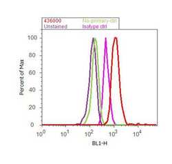
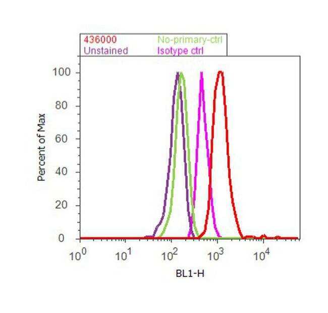
- Flow cytometry analysis of MMP-2/CLG4A was done on HT-29 cells. Cells were fixed with 70% ethanol for 10 minutes, permeabilized with 0.25% Triton™ X-100 for 20 minutes, and blocked with 5% BSA for 30 minutes at room temperature. Cells were labeled with MMP-2/CLG4A Mouse Monoclonal Antibody (Product # 436000, red histogram) or with mouse isotype control (pink histogram) at 3-5 µg/million cells in 2.5% BSA. After incubation at room temperature for 2 hours, the cells were labeled with Alexa Fluor® 488 Rabbit Anti-Mouse Secondary Antibody (Product # A-11059) at a dilution of 1:400 for 30 minutes at room temperature. The representative 10,000 cells were acquired and analyzed for each sample using an Attune® Acoustic Focusing Cytometer. The purple histogram represents unstained control cells and the green histogram represents no-primary-antibody control.
- Submitted by
- Invitrogen Antibodies (provider)
- Main image
- Experimental details
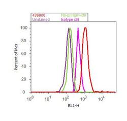
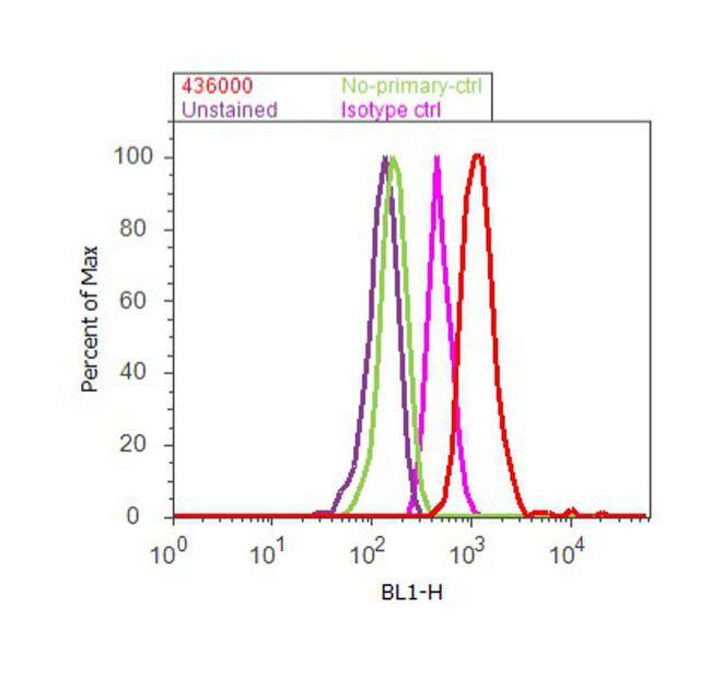
- Flow cytometry analysis of MMP-2/CLG4A was done on HT-29 cells. Cells were fixed with 70% ethanol for 10 minutes, permeabilized with 0.25% Triton™ X-100 for 20 minutes, and blocked with 5% BSA for 30 minutes at room temperature. Cells were labeled with MMP-2/CLG4A Mouse Monoclonal Antibody (Product # 436000, red histogram) or with mouse isotype control (pink histogram) at 3-5 µg/million cells in 2.5% BSA. After incubation at room temperature for 2 hours, the cells were labeled with Alexa Fluor® 488 Rabbit Anti-Mouse Secondary Antibody (Product # A-11059) at a dilution of 1:400 for 30 minutes at room temperature. The representative 10,000 cells were acquired and analyzed for each sample using an Attune® Acoustic Focusing Cytometer. The purple histogram represents unstained control cells and the green histogram represents no-primary-antibody control.
Supportive validation
- Submitted by
- Invitrogen Antibodies (provider)
- Main image
- Experimental details
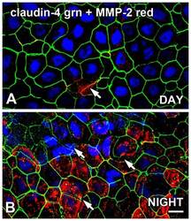
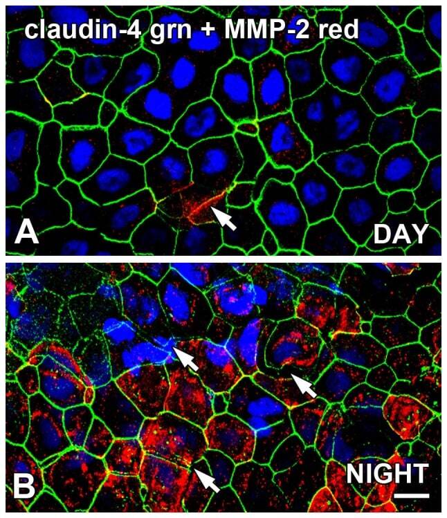
- Figure 5 Claudin-4 expression on Xenopus surface CE is disrupted at night, and is inversely associated with MMP-2 expression. Flat-mounted whole corneas were double-labeled for localization of claudin-4 and MMP-2 immunoreactivity in the late afternoon (DAY; 9 hours after lights on in a 12L:12D cycle) and in the late night (NIGHT; 3 hours before lights on). ( A ) In the late afternoon, claudin-4 (green) immunolabeling of the lateral membranes of the surface CE was generally intact, and some sporadic labeling of MMP-2 (red) was also present on the lateral membranes and/or cytoplasm (arrows) which was sometimes co-localized (yellow) with occludin. ( B ) During the late night, most of the surface CE cells exhibited intact claudin-4 (green) labeling on their lateral membranes, but there were also many clusters of surface CE cells in which the claudin-4 immunoreactivity was disrupted (arrows) and was accompanied by high levels of MMP-2 immunoreactivity. Specimens were stained with the blue nuclear DAPI stain. Scale bar = 20 um.
- Submitted by
- Invitrogen Antibodies (provider)
- Main image
- Experimental details
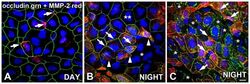
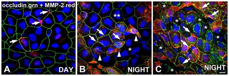
- Figure 4 Occludin expression on Xenopus surface CE is disrupted at night, and is inversely associated with MMP-2 expression. Flat-mounted whole corneas were double-labeled for localization of occludin and MMP-2 immunoreactivity in the late afternoon (DAY; 9 hours after lights on in a 12L:12D cycle) and in the late night (NIGHT; 3 hours before lights on). ( A ) In the late afternoon, occludin (green) immunolabeling of the lateral membranes of the surface CE was generally intact, and some sporadic labeling of MMP-2 (red) was also present on the lateral membranes and/or cytoplasm (arrows) which was sometimes co-localized (yellow) with occludin. ( B ) During the late night, most of the surface CE cells exhibited intact occludin (green) labeling on their lateral membranes (double asterisks), but there were also many clusters of surface CE cells that lacked occludin immunoreactivity in their lateral membranes (single asterisk). In neighboring cells of some occludin-negative clusters, there was a gap between the occludin-labeled lateral membranes, indicating the disruption of tight junctions between the cells (arrows). MMP-2 (red) was very often expressed in the cells that exhibited gaps between the lateral membranes. Also, in these areas of high MMP-2 expression, upward-folding flaps of surface cells were lifting from the CE surface, representing cells in the act of desquamation (arrowheads). ( C ) In some cell clusters that were almost devoid of occludin labeling on their lateral
- Submitted by
- Invitrogen Antibodies (provider)
- Main image
- Experimental details
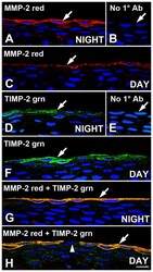
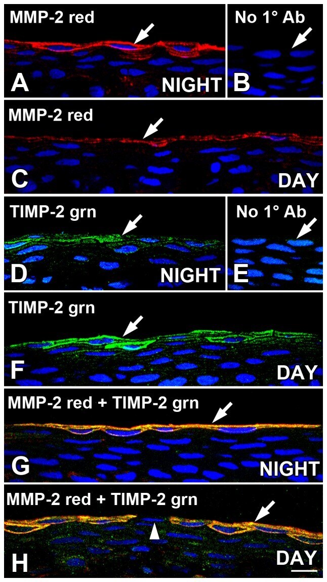
- Figure 1 MMP-2 and TIMP are co-localized in Xenopus leavis surface corneal epithelium sections. Cryostat sections of corneas obtained during the late dark period (NIGHT: two hrs before lights on) or early light period (DAY; two hrs after lights on) were immunolabeled with MMP-2 and or/TIMP-2 antibodies and analyzed with confocal microscopy. ( A ) At nighttime, MMP-2 immunoreactivity (red) was localized in cell clusters mainly to the surface layer of CE cells (arrow), with some labeling also present in the underlying sub-superficial layer of cells. Immunolabeling in the deeper layers of the CE was very sparse. ( B ) As negative controls, corneal sections obtained at night were processed for immunohistochemistry in the absence of primary antibody, and no specific immunoreactivity was observed (arrow). ( C ) At early daytime, intensity of MMP-2 immunoreactivity (red) was generally diminished relative to the night time levels (arrow). ( D ) At late nighttime, TIMP-2 immunoreactivity (green) was localized in cell clusters mainly to the surface layer of CE cells (arrow), with some labeling also present in the underlying sub-superficial layer of cells. Immunolabeling in the deeper layers of the CE was very sparse. ( E ) As negative controls, corneal sections obtained during the daytime were processed for immunohistochemistry in the absence of primary antibody, and no specific immunoreactivity was observed (arrow). ( F ) At early daytime, intensity of TIMP-2 immunoreactivity (green)
- Submitted by
- Invitrogen Antibodies (provider)
- Main image
- Experimental details
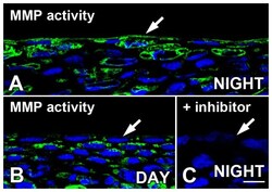
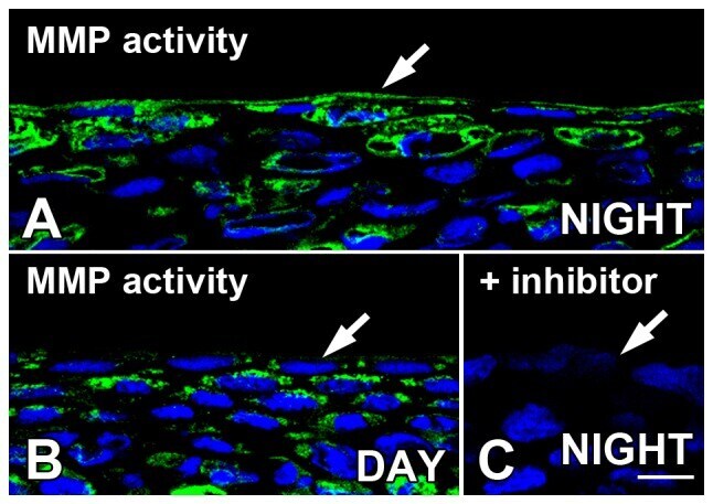
- Figure 2 In situ zymography demonstrates MMP gelatinase activity in Xenopus surface corneal epithelium. The presence of green fluorescence with confocal microscopy indicates the presence of gelatinase enzyme activity in unfixed cornea sections obtained during the late dark period (NIGHT: two hrs before lights on) or early light period (DAY; two hrs after lights on). ( A ) Late at night, MMP activity (green) was present in the surface layers of the CE (arrow) with some labeling also present in the deeper layers of the CE. In the early daytime, MMP activity (green) in the surface CE (arrow) appeared to be lower early in the light period with some labeling persisting in the deeper layers of the cornea. (C) Inclusion of a specific inhibitor of MMP-2 and MMP-9 (1.0 um phenanthroline) in the incubation mixture blocked most of the gelatinase activity. Sections were stained with DAPI, which stained the nuclei blue. Scale bar = 20 um.
- Submitted by
- Invitrogen Antibodies (provider)
- Main image

- Experimental details
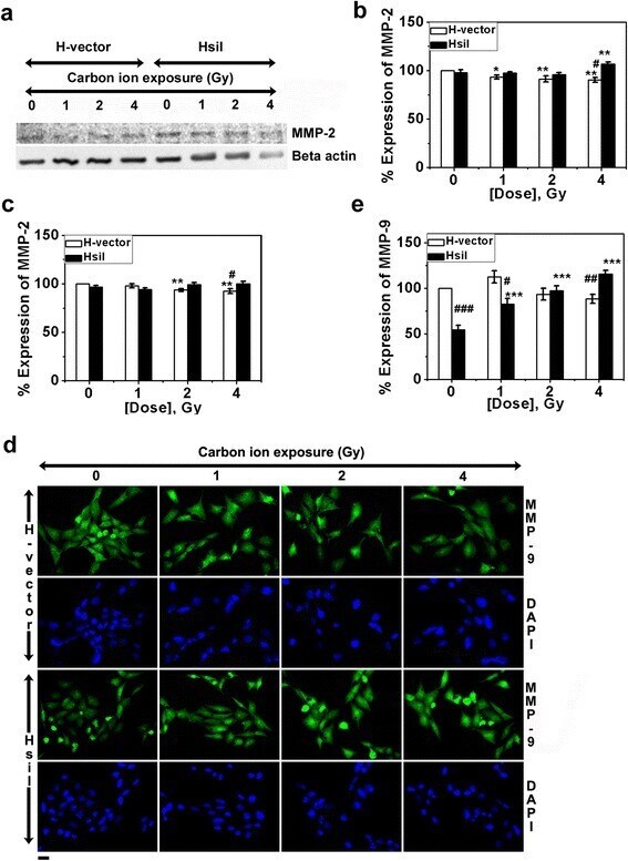
- Fig. 3 Expression of MMP-2 and MMP-9 protein after carbon ion exposure (0- 4 Gy) in H-vector and HsiI cells. a Typical western blot of MMP-2 and beta actin (used as internal control). b Band intensities of western blot of MMP-2 were measured by ImageJ software and normalized with respect to beta actin and plotted as % expression of MMP-2 considering MMP-2 expression in un-irradiated H-vector as 100 %. c Expression of MMP-2 was also measured by IF technique. Intensities of the immunostained cells for MMP-2 were measured by ImageJ software, normalized with respect to beta actin and plotted as mentioned above. Typical immunostained cells are shown in Additional file 2 . d Expression of MMP-9 was measured by IF technique and typical immunostained cells are shown here. Scale bar represents 20 mum. e Intensities of the immunostained cells for MMP-9 were measured by ImageJ software, normalized with respect to beta actin and plotted as mentioned above. Each bar diagram represents the mean +- SD of three independent experiments
- Submitted by
- Invitrogen Antibodies (provider)
- Main image
- Experimental details
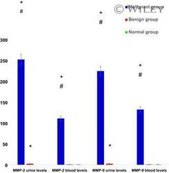
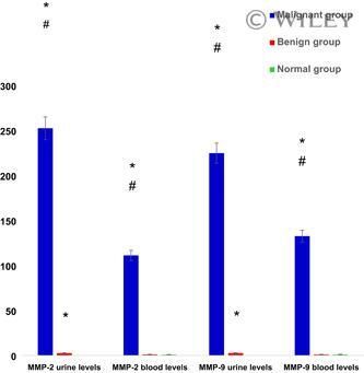
- 1 Blood and urinary protein levels of MMP-2 and MMP-9 in groups of the study by Western blot. *Significant P versus control group, # significant P versus benign bladder disorders group. Y axis represents a ratio between each target gene expression and housekeeping gene expression. MMP, matrix metalloproteinase
- Submitted by
- Invitrogen Antibodies (provider)
- Main image

- Experimental details
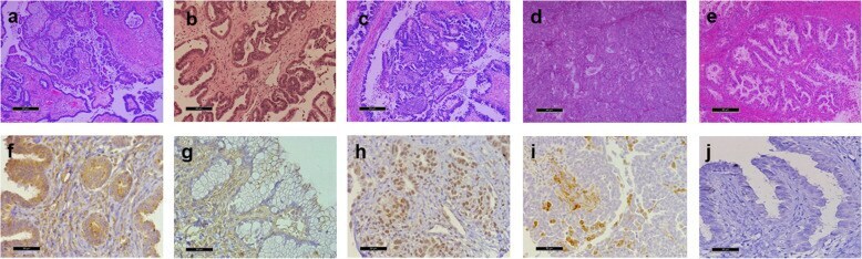
- Fig. 1 Representative hematoxilin-eosin stained sections. a Serous borderline tumor, b Low grade serous carcinoma, c High grade serous carcinoma, d Endometrioid carcinoma, and e Mucinous carcinoma. Immunohistochemistry for MMP-2 and MMP-9 in epithelial ovarian tumors. f MMP-2 in epithelium and stroma, of endometrioid tumor. g MMP-2 in stroma of mucinous tumor. h MMP-9 in epithelium of high grade serous carcinoma. i MMP-9 in stroma of endometrioid carcinoma. j negative control. Bars represents 50 mum
- Submitted by
- Invitrogen Antibodies (provider)
- Main image
- Experimental details
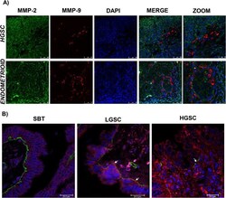
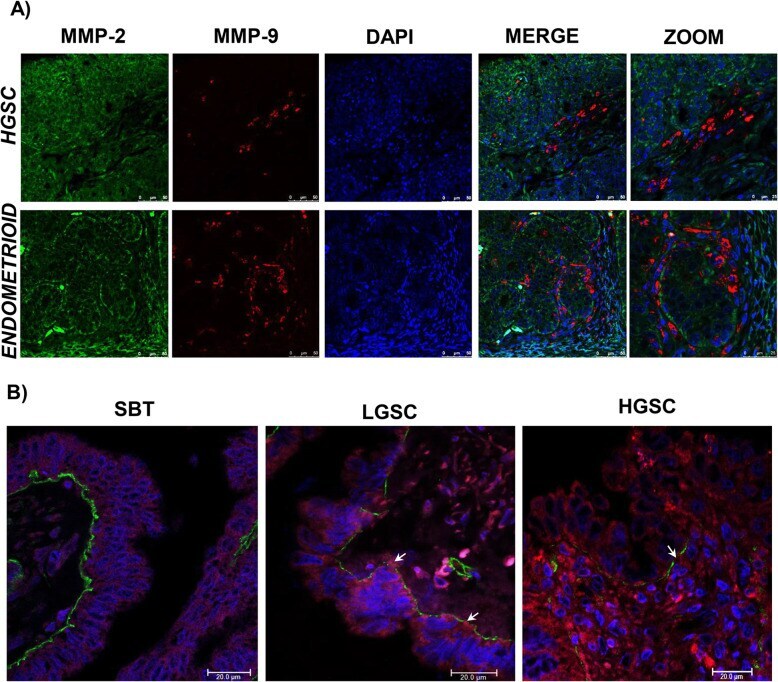
- Fig. 2 a Immunofluorescence for high grade serous carcinoma (HGSC) and endometrioid carcinoma. MMPs proteins were observed in the cytoplasm stained using MMP-2 antibody (green) and MMP-9 antibody (red), nuclei were stained with DAPI (blue). MMPs are shown in separate locations within the tumor. b Immunofluorescence for serous borderline tumor, low grade serous carcinoma (LGSC), and high grade serous carcinoma (HGSC). MMP-9 antibody (red) evidenced the cytoplasm of epithelial tumor cells, while the basal lamina was stained with collagen IV antibody (green) and the nuclei using DAPI (blue). Basal lamina discontinuities were observed in relation to MMP presence (arrows), mainly in LGSC
- Submitted by
- Invitrogen Antibodies (provider)
- Main image
- Experimental details
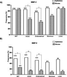
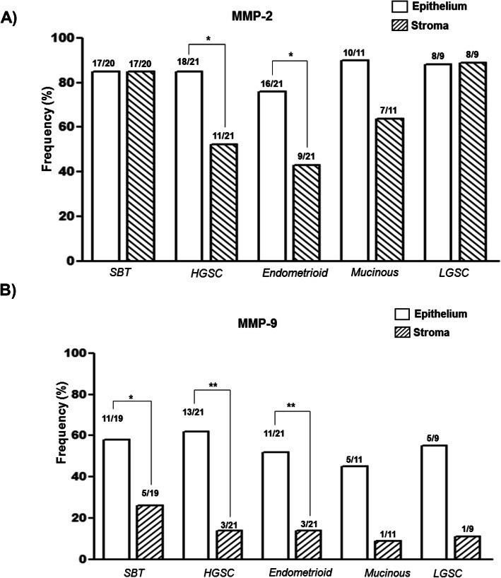
- Fig. 3 Frequency of expression of MMP-2 ( a ) and MMP-9 ( b ) in ovarian tumors. Bars represent the proportions of positive immunohistochemical reaction in epithelium and the stroma of tissue samples. Statistical analysis was performed using the test for contrasting the proportions of the groups,* P
- Submitted by
- Invitrogen Antibodies (provider)
- Main image
- Experimental details
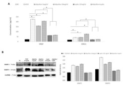
- Figure 5 AdipoRon and leptin exert antagonizing effects on HIF-1alpha, CXCL1, VEGF-A, MMP-2 and MMP-9 protein levels. ( A ) Elisa assay of CXCL1 and VEGF-A; ( B ) representative images of immunoblotting of total MMP-2 and MMP-9 proteins and their quantitation. HUVEC endothelial cells were treated with AdipoRon (5 or 10 mug/mL), Leptin (125 ng/mL) and a combination of AdipoRon+leptin (5 mug/mL and 125 ng/mL, respectively) for 24 h. Untreated cells were used as negative control (NC); VEGF (10 ng/mL) was used as a positive control. Data are reported as mean +- Standard Deviation (SD) of three independent experiments were reported. * p < 0.05.
