Explore
 Validate
 Learn
Learn Western blot
 Immunocytochemistry
Antibody data
- Antibody Data
- Antigen structure
- References [0]
- Comments [0]
- Validations
- Western blot [2]
- Immunohistochemistry [2]
- Other assay [1]
Submit
Validation data
Reference
Comment
Report error
- Product number
- PA5-16851 - Provider product page

- Provider
- Invitrogen Antibodies
- Product name
- Anti-MMP9 Polyclonal Antibody
- Antibody type
- Polyclonal
- Antigen
- Synthetic peptide
- Description
- PA5-16851 targets MMP-9 (92kDa Collagenase IV) in IHC (P) and WB applications and shows reactivity with Guinea Pig and Human samples. The PA5-16851 immunogen is a synthetic peptide derived from near C-terminal of human MMP-9 protein.
- Reactivity
- Human, Guinea Pig
- Host
- Rabbit
- Isotype
- IgG
- Vial size
- 500 µL
- Concentration
- Lot Dependent
- Storage
- Store at 4°C short term. For long term storage, store at -20°C, avoiding freeze/thaw cycles.
No comments: Submit comment
Supportive validation
- Submitted by
- Invitrogen Antibodies (provider)
- Main image

- Experimental details
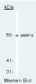
- Western blot of MMP-9 (92kDa Collagenase IV) using MMP-9 (92kDa Collagenase IV) Polyclonal Antibody (Product # PA5-16851) on HFL-1+TPA med Cells.
- Submitted by
- Invitrogen Antibodies (provider)
- Main image

- Experimental details
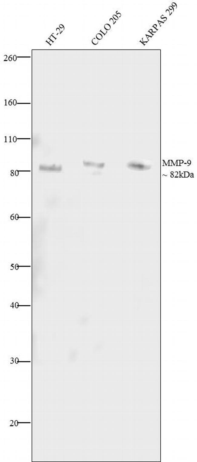
- Western blot analysis was performed on conditioned media of serum starved HT-29 (Lane 1), serum starved COLO 205 (Lane 2) and serum starved KARPAS 299 (Lane 3). The blots were probed with Anti-MMP-9 Rabbit Polyclonal Antibody (Product # PA5-16851, 2 µg/mL) and detected by chemiluminescence using Goat anti-Rabbit IgG (H+L) Superclonal™ Secondary Antibody, HRP conjµgate (Product # A27036, 0.4µg/mL, 1:2500 dilution). A ~ 82 kDa band corresponding to MMP-9 was observed across cell lines tested. MMP9 is secreted as a 92 kDa zymogen. Cleavage of ProMMP-9 results in the active enzyme, having a molecular weight of approximately 82 kDa. Known quantity of protein samples were electrophoresed using Novex® NuPAGE® 10 % Bis-Tris gel (Product # NP0302BOX), XCell SureLock™ Electrophoresis System (Product # EI0002) and Novex® Sharp Pre-Stained Protein Standard (Product # LC5800). Resolved proteins were then transferred onto a nitrocellulose membrane by overnight transfer method. The membrane was probed with the relevant primary and secondary Antibody following blocking with 5 % skimmed milk. Chemiluminescent detection was performed using Pierce™ ECL Western Blotting Substrate (Product # 32106).
Supportive validation
- Submitted by
- Invitrogen Antibodies (provider)
- Main image

- Experimental details
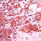
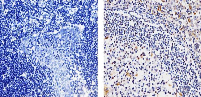
- Formalin-fixed, paraffin-embedded human placenta stained with MMP-9 antibody2 using peroxidase-conjugate and AEC chromogen. Note membrane staining of trophoblasts.
- Submitted by
- Invitrogen Antibodies (provider)
- Main image
- Experimental details
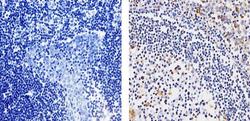
- Immunohistochemistry analysis of MMP-9 (92kDa Collagenase IV) showing staining in the cytoplasm of paraffin-embedded human tonsil tissue (right) compared to a negative control without primary antibody (left). To expose target proteins, antigen retrieval was performed using 10mM sodium citrate (pH 6.0), microwaved for 8-15 min. Following antigen retrieval, tissues were blocked in 3% H2O2-methanol for 15 min at room temperature, washed with ddH2O and PBS, and then probed with a MMP-9 (92kDa Collagenase IV) Rabbit Polyclonal Antibody (Product # PA5-16851) diluted in 3% BSA-PBS at a dilution of 1:100 for 1 hour at 37ºC in a humidified chamber. Tissues were washed extensively in PBST and detection was performed using an HRP-conjugated secondary antibody followed by colorimetric detection using a DAB kit. Tissues were counterstained with hematoxylin and dehydrated with ethanol and xylene to prep for mounting.
Supportive validation
- Submitted by
- Invitrogen Antibodies (provider)
- Main image

- Experimental details
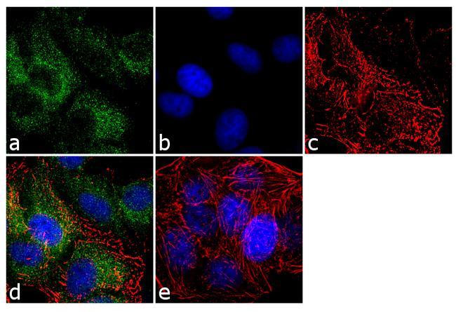
- Immunofluorescence analysis of MMP-9 was performed using 70% confluent log phase MCF-7 cells. The cells were fixed with 4% paraformaldehyde for 10 minutes, permeabilized with 0.1% Triton™ X-100 for 10 minutes, and blocked with 1% BSA for 1 hour at room temperature. The cells were labeled with MMP-9 (92 kDa Collagenase IV) Rabbit Polyclonal Antibody (Product # PA5-16851) at 2µg/mL in 0.1% BSA and incubated for 3 hours at room temperature and then labeled with Goat anti-Rabbit IgG (H+L) Superclonal™ Secondary Antibody, Alexa Fluor® 488 conjµgate (Product # A27034) at a dilution of 1:2000 for 45 minutes at room temperature (Panel a: green). Nuclei (Panel b: blue) were stained with SlowFade® Gold Antifade Mountant with DAPI (Product # S36938). F-actin (Panel c: red) was stained with Alexa Fluor® 555 Rhodamine Phalloidin (Product # R415, 1:300). Panel d represents the merged image showing cytoplasmic localization. Panel e shows the no primary antibody control. The images were captured at 60X magnification.