Explore
 Validate
 Learn
Learn Western blot
 Immunocytochemistry
Antibody data
- Antibody Data
- Antigen structure
- References [2]
- Comments [0]
- Validations
- Immunocytochemistry [1]
- Immunohistochemistry [1]
- Other assay [1]
Submit
Validation data
Reference
Comment
Report error
- Product number
- PA5-27191 - Provider product page

- Provider
- Invitrogen Antibodies
- Product name
- MMP9 Polyclonal Antibody
- Antibody type
- Polyclonal
- Antigen
- Recombinant full-length protein
- Description
- Recommended positive controls: MCF-7 (starvation, 200 nM PMA treatment for 24 hr, and 5 µg/mL Brefeldin A treatment for 14.5 hr), Huh7. Predicted reactivity: Mouse (82%), Rat (84%), Dog (81%), Pig (84%), Rabbit (89%), Rhesus Monkey (96%), Bovine (85%). Store product as a concentrated solution. Centrifuge briefly prior to opening the vial.
- Reactivity
- Human, Mouse, Bovine
- Host
- Rabbit
- Isotype
- IgG
- Vial size
- 100 μL
- Concentration
- 1.96 mg/mL
- Storage
- Store at 4°C short term. For long term storage, store at -20°C, avoiding freeze/thaw cycles.
Submitted references HDAC1 promotes the migration of human myeloma cells via regulation of the lncRNA/Slug axis.
Autophagy Induced by Palmitic Acid Regulates Neutrophil Adhesion Through the Granule-Dependent Degradation of αMβ2 Integrin in Dairy Cows With Fatty Liver.
Zheng L, Zhang A, Liu J, Liu M, Zhang Y
International journal of molecular medicine 2022 Jan;49(1)
International journal of molecular medicine 2022 Jan;49(1)
Autophagy Induced by Palmitic Acid Regulates Neutrophil Adhesion Through the Granule-Dependent Degradation of αMβ2 Integrin in Dairy Cows With Fatty Liver.
Peng Z, Zhao C, Du X, Yang Y, Li Y, Song Y, Fang B, Zhang Y, Qin X, Zhang Y, Li X, Wang Z, Li X, Liu G
Frontiers in immunology 2021;12:726829
Frontiers in immunology 2021;12:726829
No comments: Submit comment
Supportive validation
- Submitted by
- Invitrogen Antibodies (provider)
- Main image

- Experimental details
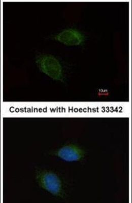
- Immunofluorescent analysis of MMP9 in methanol-fixed HeLa cells using a MMP9 polyclonal antibody (Product # PA5-27191) at a 1:200 dilution.
Supportive validation
- Submitted by
- Invitrogen Antibodies (provider)
- Main image

- Experimental details
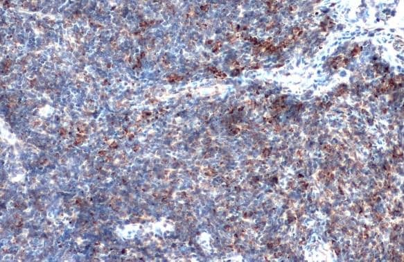
- Immunohistochemistry (Paraffin) analysis of MMP9 was performed in paraffin-embedded mouse lymph node tissue using MMP9 Polyclonal Antibody (Product # PA5-27191) at a dilution of 1:500. Antigen Retrieval: Citrate buffer, pH 6.0, 15 min.
Supportive validation
- Submitted by
- Invitrogen Antibodies (provider)
- Main image

- Experimental details
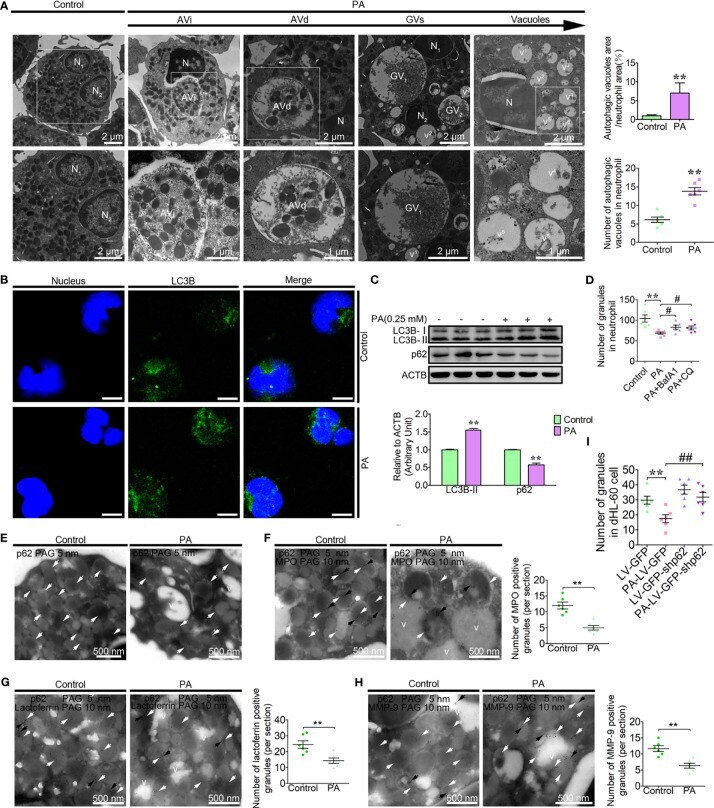
- Figure 2 PA triggers autophagy-dependent degradation of the granules and vacuolation in dairy cow neutrophils. (A) Representative transmission electron micrographs of control and PA (0.25 mM)-treated neutrophils (left). White arrows indicate autophagic vacuoles (AVi, AVd, glycogen vacuoles, and vacuoles). N (N1, N2, N3...), nucleus. Scale bars as indicated. The area ratio of autophagic vacuoles to neutrophils and the number of autophagic vacuoles in neutrophils were determined (right, n = 6). Data represent the mean +- s.e.m. (** p < 0.01 versus the control group; significance calculated using t -test). (B) Autophagy levels were evaluated using confocal microscopy in control and PA-treated neutrophils. Neutrophils were stained with anti-LC3B antibody and nuclear DNA was stained with Hoechst 33258. Scale bar, 5 mum. (C) Immunoblot for LC3B and p62 in control and PA-treated bovine neutrophils. ACTB was used as a loading control ( n = 3). Data represent the mean +- s.e.m. (** p < 0.01 versus the control group; significance calculated using two-way ANOVA). (D) The number of granules in PA-treated neutrophils (autophagy pathway was blocked using BafA1, CQ or not, n = 6). Data represent the mean +- s.e.m. (** p < 0.01 versus the control group, # < 0.05 and versus the PA-treated group; Significance calculated using one-way ANOVA). (E) Immunogold electron micrograph showing the localization of p62 in control and PA-treated bovine neutrophils. (F) P62-mediated PA-triggered autophagy-d