Explore
 Validate
 Learn
Learn Western blot
 Immunocytochemistry
Antibody data
- Antibody Data
- Antigen structure
- References [4]
- Comments [0]
- Validations
- Immunocytochemistry [1]
- Immunohistochemistry [1]
Submit
Validation data
Reference
Comment
Report error
- Product number
- HPA002834 - Provider product page

- Provider
- Atlas Antibodies
- Proper citation
- Atlas Antibodies Cat#HPA002834, RRID:AB_1079711
- Product name
- Anti-PTGS1
- Antibody type
- Polyclonal
- Description
- Polyclonal Antibody against Human PTGS1, Gene description: prostaglandin-endoperoxide synthase 1 (prostaglandin G/H synthase and cyclooxygenase), Alternative Gene Names: COX1, PGHS-1, PTGHS, Validated applications: ICC, IHC, WB, Uniprot ID: P23219, Storage: Store at +4°C for short term storage. Long time storage is recommended at -20°C.
- Reactivity
- Human
- Host
- Rabbit
- Conjugate
- Unconjugated
- Isotype
- IgG
- Vial size
- 100 µl
- Concentration
- 0.1 mg/ml
- Storage
- Store at +4°C for short term storage. Long time storage is recommended at -20°C.
- Handling
- The antibody solution should be gently mixed before use.
Submitted references Plasma proteins elevated in severe asthma despite oral steroid use and unrelated to Type-2 inflammation
Cytoplasmic PPARγ is a marker of poor prognosis in patients with Cox-1 negative primary breast cancers
Understanding the Interplay between COX-2 and hTERT in Colorectal Cancer Using a Multi-Omics Analysis
Expression profiling of microdissected cell populations selected from basal cells in normal epidermis and basal cell carcinoma
Sparreman Mikus M, Kolmert J, Andersson L, Östling J, Knowles R, Gómez C, Ericsson M, Thörngren J, Emami Khoonsari P, Dahlén B, Kupczyk M, De Meulder B, Auffray C, Bakke P, Beghe B, Bel E, Caruso M, Chanez P, Chawes B, Fowler S, Gaga M, Geiser T, Gjomarkaj M, Horváth I, Howarth P, Johnston S, Joos G, Krug N, Montuschi P, Musial J, Niżankowska-Mogilnicka E, Olsson H, Papi A, Rabe K, Sandström T, Shaw D, Siafakas N, Uhlén M, Riley J, Bates S, Middelveld R, Wheelock C, Chung K, Adcock I, Sterk P, Djukanovic R, Nilsson P, Dahlén S, James A
European Respiratory Journal 2022;59(2):2100142
European Respiratory Journal 2022;59(2):2100142
Cytoplasmic PPARγ is a marker of poor prognosis in patients with Cox-1 negative primary breast cancers
Shao W, Kuhn C, Mayr D, Ditsch N, Kailuwait M, Wolf V, Harbeck N, Mahner S, Jeschke U, Cavaillès V, Sixou S
Journal of Translational Medicine 2020;18(1)
Journal of Translational Medicine 2020;18(1)
Understanding the Interplay between COX-2 and hTERT in Colorectal Cancer Using a Multi-Omics Analysis
Ayiomamitis G, Notas G, Vasilakaki T, Tsavari A, Vederaki S, Theodosopoulos T, Kouroumalis E, Zaravinos A
Cancers 2019;11(10):1536
Cancers 2019;11(10):1536
Expression profiling of microdissected cell populations selected from basal cells in normal epidermis and basal cell carcinoma
Asplund A, Gry Björklund M, Sundquist C, Strömberg S, Edlund K, Östman A, Nilsson P, Pontén F, Lundeberg J
British Journal of Dermatology 2008;158(3):527-538
British Journal of Dermatology 2008;158(3):527-538
No comments: Submit comment
Supportive validation
- Submitted by
- Atlas Antibodies (provider)
- Main image

- Experimental details
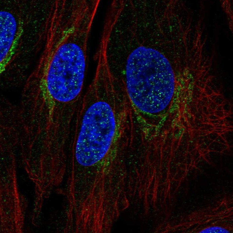
- Immunofluorescent staining of human cell line BJ shows localization to the Golgi apparatus.
- Sample type
- Human
Supportive validation
- Submitted by
- Atlas Antibodies (provider)
- Enhanced method
- Orthogonal validation
- Main image

- Experimental details
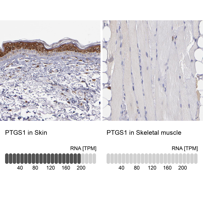
- Immunohistochemistry analysis in human skin and skeletal muscle tissues using Anti-PTGS1 antibody. Corresponding PTGS1 RNA-seq data are presented for the same tissues.
- Sample type
- Human
- Protocol
- Protocol