Explore
 Validate
 Learn
Learn Western blot
 Immunocytochemistry
Antibody data
- Antibody Data
- Antigen structure
- References [5]
- Comments [0]
- Validations
- Western blot [1]
- Flow cytometry [1]
Submit
Validation data
Reference
Comment
Report error
- Product number
- AF4198 - Provider product page

- Provider
- Novus Biologicals
- Product name
- Goat Polyclonal COX-2 Antibody
- Antibody type
- Polyclonal
- Description
- Immunogen affinity purified. Detects human and mouse COX-2 in Western blots. In Western blots, less than 1% cross-reactivity with recombinant human COX-1 is observed.
- Reactivity
- Human, Mouse
- Host
- Goat
- Isotype
- IgG
- Vial size
- 100 ug
- Concentration
- LYOPH
- Storage
- Use a manual defrost freezer and avoid repeated freeze-thaw cycles. 12 months from date of receipt, -20 to -70 degreesC as supplied. 1 month, 2 to 8 degreesC under sterile conditions after reconstitution. 6 months, -20 to -70 degreesC under sterile conditions after reconstitution.
Submitted references Increased Toxoplasma gondii Intracellular Proliferation in Human Extravillous Trophoblast Cells (HTR8/SVneo Line) Is Sequentially Triggered by MIF, ERK1/2, and COX-2.
Multi-Staged Regulation of Lipid Signaling Mediators during Myogenesis by COX-1/2 Pathways.
TET2 Regulates the Neuroinflammatory Response in Microglia.
Response gene to complement 32 expression in macrophages augments paracrine stimulation-mediated colon cancer progression.
Stromal versus tumoral inflammation differentially contribute to metastasis and poor survival in laryngeal squamous cell carcinoma.
Milian ICB, Silva RJ, Manzan-Martins C, Barbosa BF, Guirelli PM, Ribeiro M, de Oliveira Gomes A, Ietta F, Mineo JR, Silva Franco P, Ferro EAV
Frontiers in microbiology 2019;10:852
Frontiers in microbiology 2019;10:852
Multi-Staged Regulation of Lipid Signaling Mediators during Myogenesis by COX-1/2 Pathways.
Mo C, Wang Z, Bonewald L, Brotto M
International journal of molecular sciences 2019 Sep 4;20(18)
International journal of molecular sciences 2019 Sep 4;20(18)
TET2 Regulates the Neuroinflammatory Response in Microglia.
Carrillo-Jimenez A, Deniz Ö, Niklison-Chirou MV, Ruiz R, Bezerra-Salomão K, Stratoulias V, Amouroux R, Yip PK, Vilalta A, Cheray M, Scott-Egerton AM, Rivas E, Tayara K, García-Domínguez I, Garcia-Revilla J, Fernandez-Martin JC, Espinosa-Oliva AM, Shen X, St George-Hyslop P, Brown GC, Hajkova P, Joseph B, Venero JL, Branco MR, Burguillos MA
Cell reports 2019 Oct 15;29(3):697-713.e8
Cell reports 2019 Oct 15;29(3):697-713.e8
Response gene to complement 32 expression in macrophages augments paracrine stimulation-mediated colon cancer progression.
Zhao P, Wang B, Zhang Z, Zhang W, Liu Y
Cell death & disease 2019 Oct 10;10(10):776
Cell death & disease 2019 Oct 10;10(10):776
Stromal versus tumoral inflammation differentially contribute to metastasis and poor survival in laryngeal squamous cell carcinoma.
Höing B, Kanaan O, Altenhoff P, Petri R, Thangavelu K, Schlüter A, Lang S, Bankfalvi A, Brandau S
Oncotarget 2018 Feb 2;9(9):8415-8426
Oncotarget 2018 Feb 2;9(9):8415-8426
No comments: Submit comment
Supportive validation
- Submitted by
- Novus Biologicals (provider)
- Main image
- Experimental details
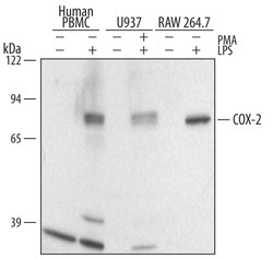
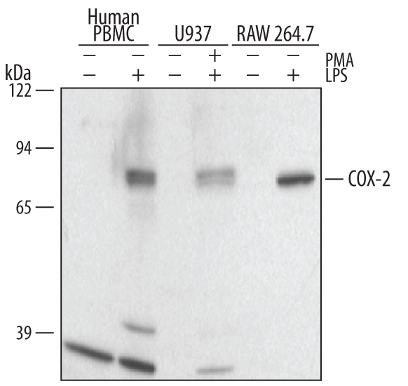
- Detection of Human and Mouse COX-2 by Western Blot. Western blot shows lysates of human peripheral blood mononuclear cell (PBMC) and RAW 264.7 mouse monocyte/macrophage cell line untreated (-) or treated (+) with 1 ug/mL LPS for 24 hours and U937 human histiocytic lymphoma cell line untreated or treated with 100 nM PMA and 1 ug/mL LPS for 48 hours and 24 hours, respectively. PVDF membrane was probed with 1 µg/mL of Human/Mouse COX-2 Polyclonal Antibody (Catalog # AF4198), followed by HRP-conjugated Anti-Goat IgG Secondary Antibody (Catalog # HAF109). A specific band was detected for COX-2 at approximately 75 kDa (as indicated). This experiment was conducted under reducing conditions and using Immunoblot Buffer Group 2.
Supportive validation
- Submitted by
- Novus Biologicals (provider)
- Main image
- Experimental details
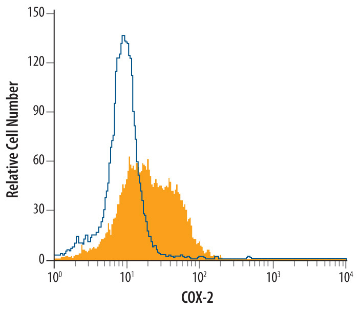
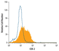
- Detection of COX-2 in RAW 264.7 Mouse Cell Line by Flow Cytometry. RAW 264.7 mouse monocyte/macrophage cell line treated with 1 μg/mL LPS for 24 hours was stained with Goat Anti-Human/Mouse COX-2 Antigen Affinity-purified Polyclonal Antibody (Catalog # AF4198, filled histogram) or control antibody (Catalog # AB-108-C, open histogram), followed by Allophycocyanin-conjugated Anti-Goat IgG Secondary Antibody (Catalog # F0108). To facilitate intracellular staining, cells were fixed with paraformaldehyde and permeabilized with saponin.