 Explore
 Validate
 Learn
Learn Western blot
 Immunocytochemistry
Antibody data
- Antibody Data
- Antigen structure
- References [2]
- Comments [0]
- Validations
- Western blot [1]
- Immunohistochemistry [1]
Submit
Validation data
Reference
Comment
Report error
- Product number
- MAB4198 - Provider product page

- Provider
- Novus Biologicals
- Product name
- Mouse Monoclonal COX-2 Antibody
- Antibody type
- Monoclonal
- Description
- Protein A or G purified from hybridoma culture supernatant. Detects human COX-2 in Western blots.
- Reactivity
- Human
- Host
- Mouse
- Conjugate
- Unconjugated
- Isotype
- IgG
- Vial size
- 100 ug
- Concentration
- LYOPH
- Storage
- Use a manual defrost freezer and avoid repeated freeze-thaw cycles. 12 months from date of receipt, -20 to -70 degreesC as supplied. 1 month, 2 to 8 degreesC under sterile conditions after reconstitution. 6 months, -20 to -70 degreesC under sterile conditions after reconstitution.
Submitted references Evaluation of an Aqueous Extract from Horseradish Root (Armoracia rusticana Radix) against Lipopolysaccharide-Induced Cellular Inflammation Reaction.
Adipose-derived mesenchymal stem cells modulate CD14(++)CD16(+) expression on monocytes from sepsis patients in vitro via prostaglandin E2.
Herz C, Tran HT, Márton MR, Maul R, Baldermann S, Schreiner M, Lamy E
Evidence-based complementary and alternative medicine : eCAM 2017;2017:1950692
Evidence-based complementary and alternative medicine : eCAM 2017;2017:1950692
Adipose-derived mesenchymal stem cells modulate CD14(++)CD16(+) expression on monocytes from sepsis patients in vitro via prostaglandin E2.
Qiu G, Zheng G, Ge M, Huang L, Tong H, Chen P, Lai D, Hu Y, Cheng B, Shu Q, Xu J
Stem cell research & therapy 2017 Apr 26;8(1):97
Stem cell research & therapy 2017 Apr 26;8(1):97
No comments: Submit comment
Supportive validation
- Submitted by
- Novus Biologicals (provider)
- Main image

- Experimental details
- Detection of Human COX-2 by Western Blot. Western blot shows lysates of human peripheral blood mononuclear cells (PBMC) untreated (-) or treated (+) with LPS. PVDF membrane was probed with 1 µg/mL of Mouse Anti-Human COX-2 Monoclonal Antibody (Catalog # MAB4198), followed by HRP-conjugated Anti-Mouse IgG Secondary Antibody (Catalog # HAF007). A specific band was detected for COX-2 at approximately 75 kDa (as indicated). This experiment was conducted under reducing conditions and using Immunoblot Buffer Group 2.

Supportive validation
- Submitted by
- Novus Biologicals (provider)
- Main image
- Experimental details
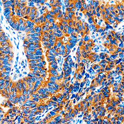
- COX-2 in Human Breast Cancer Tissue. COX-2 was detected in immersion fixed paraffin-embedded sections of human breast cancer tissue using Mouse Anti-Human COX-2 Monoclonal Antibody (Catalog # MAB4198) at 1.7 µg/mL overnight at 4 °C. Tissue was stained using the Anti-Mouse HRP-DAB Cell & Tissue Staining Kit (brown; Catalog # CTS002) and counterstained with hematoxylin (blue). Specific staining was localized to cytoplasm. View our protocol for Chromogenic IHC Staining of Paraffin-embedded Tissue Sections.
