Explore
 Validate
 Learn
Learn Western blot
 Immunocytochemistry
Antibody data
- Antibody Data
- Antigen structure
- References [5]
- Comments [0]
- Validations
- Immunocytochemistry [2]
- Immunohistochemistry [1]
- Other assay [3]
Submit
Validation data
Reference
Comment
Report error
- Product number
- PA5-17614 - Provider product page

- Provider
- Invitrogen Antibodies
- Product name
- COX2 Polyclonal Antibody
- Antibody type
- Polyclonal
- Antigen
- Synthetic peptide
- Description
- Antibodies to this protein (and modification) were previously sold as part of a Thermo Scientific Cellomics High Content Screening Kit. This replacement antibody is now recommended for researchers who need an antibody for high content cell based assays. It has been thoroughly tested and validated for cellular immunofluorescence (IF) applications. Further optimization including the selection of the most appropriate fluorescent Dylight conjugated secondary antibody may have to be performed for your high content assay. It is not recommended to aliquot this antibody.
- Reactivity
- Human, Mouse
- Host
- Rabbit
- Isotype
- IgG
- Vial size
- 100 µL
- Concentration
- 28 µg/mL
- Storage
- -20°C
Submitted references Effects of Visfatin on Intracellular Mechanics and Catabolism in Human Primary Chondrocytes through Glycogen Synthase Kinase 3β Inactivation.
Protective Effects of Foam Rolling against Inflammation and Notexin Induced Muscle Damage in Rats.
Effect of Metformin on Development of Tendinopathy Due to Mechanical Overloading in an Animal Model.
Clinopodium vulgare L. (wild basil) extract and its active constituents modulate cyclooxygenase-2 expression in neutrophils.
Wnt signaling controls radiosensitivity via cyclooxygenase-2-mediated Ku expression in head and neck cancer.
Chang SF, Huang KC, Lee KH, Chiang YC, Lee WR, Hsieh RZ, Su YP, Wu SC
International journal of molecular sciences 2021 Jul 28;22(15)
International journal of molecular sciences 2021 Jul 28;22(15)
Protective Effects of Foam Rolling against Inflammation and Notexin Induced Muscle Damage in Rats.
Pablos A, Ceca D, Jorda A, Rivera P, Colmena C, Elvira L, Martínez-Arnau FM, Valles SL
International journal of medical sciences 2020;17(1):71-81
International journal of medical sciences 2020;17(1):71-81
Effect of Metformin on Development of Tendinopathy Due to Mechanical Overloading in an Animal Model.
Zhang J, Li F, Nie D, Onishi K, Hogan MV, Wang JH
Foot & ankle international 2020 Dec;41(12):1455-1465
Foot & ankle international 2020 Dec;41(12):1455-1465
Clinopodium vulgare L. (wild basil) extract and its active constituents modulate cyclooxygenase-2 expression in neutrophils.
Amirova KM, Dimitrova P, Marchev AS, Aneva IY, Georgiev MI
Food and chemical toxicology : an international journal published for the British Industrial Biological Research Association 2019 Feb;124:1-9
Food and chemical toxicology : an international journal published for the British Industrial Biological Research Association 2019 Feb;124:1-9
Wnt signaling controls radiosensitivity via cyclooxygenase-2-mediated Ku expression in head and neck cancer.
Chang HW, Roh JL, Jeong EJ, Lee SW, Kim SW, Choi SH, Park SK, Kim SY
International journal of cancer 2008 Jan 1;122(1):100-7
International journal of cancer 2008 Jan 1;122(1):100-7
No comments: Submit comment
Supportive validation
- Submitted by
- Invitrogen Antibodies (provider)
- Main image
- Experimental details
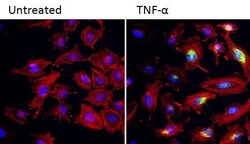
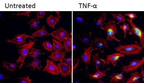
- Immunofluorescent analysis of COX-2 (green) in HeLa cells either left untreated (left lane) or treated with 100 ng/mL TNF-alpha for 20 hours. Formalin fixed cells were permeabilized with 0.1% Triton X-100 in TBS for 10 minutes at room temperature and blocked with 1% Blocker BSA (Product # 37525) for 15 minutes at room temperature. Cells were probed with a COX-2 polyclonal antibody (Product # PA5-17614) at a dilution of 1:50 for at least 1 hour at room temperature, washed with PBS, and incubated with DyLight 488 goat anti-rabbit IgG secondary antibody (Product # 35552) at a dilution of 1:400 for 30 minutes at room temperature. F-Actin (red) was stained with Dylight 554 Phalloidin (Product # 21834) and nuclei (blue) were stained with Hoechst 33342 dye (Product # 62249). Images were taken on a Thermo Scientific ArrayScan or a ToxInsight Instrument at 20X magnification.
- Submitted by
- Invitrogen Antibodies (provider)
- Main image
- Experimental details
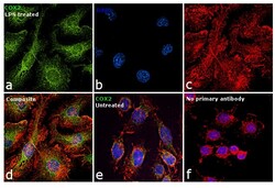
- Immunofluorescence analysis of COX2 was performed using RAW 264.7 cells treated with LPS (1 µg/mL for 24h). The cells were fixed with 4% paraformaldehyde for 10 minutes, permeabilized with 0.1% Triton™ X-100 for 15 minutes, and blocked with 1% BSA for 1 hour at room temperature. The cells were labeled with COX2 Polyclonal Antibody (Product # PA5-17614) at 1:100 dilution in 0.1% BSA, incubated at 4 degree Celsius overnight and then labeled with Goat anti-Rabbit IgG (H+L) Superclonal™ Secondary Antibody, Alexa Fluor® 488 conjugate (Product # A27034) at a dilution of 1:2000 for 45 minutes at room temperature (Panel a: green). Nuclei (Panel b: blue) were stained with ProLong™ Diamond Antifade Mountant with DAPI (Product # P36962). F-actin (Panel c: red) was stained with Rhodamine Phalloidin (Product # R415). Panel d represents the merged image showing cytoplasmic localization. Panel e represents untreated cells with reduced expression of COX2. Panel f shows cells without the primary antibody to assess background. The images were captured at 60X magnification.

Supportive validation
- Submitted by
- Invitrogen Antibodies (provider)
- Main image
- Experimental details
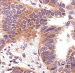
- Immunohistochemistry was performed on paraffin-embedded human lung carcinoma tissue. To expose target proteins heat induced antigen retrieval was performed using 10mM sodium citrate (pH 6.0) buffer at 95C for 10 minutes. Following antigen retrieval tissues were incubated in 3% hydrogen peroxide, blocked in 5% normal goat serum in TBST for 1 hour, and then probed with a COX2 polyclonal antibody (Product # PA5-17614) at a dilution of 1:200 overnight at 4C. Detection was performed using a peroxidase conjugated secondary reagent followed by colorimetric detection using DAB.

Supportive validation
- Submitted by
- Invitrogen Antibodies (provider)
- Main image
- Experimental details
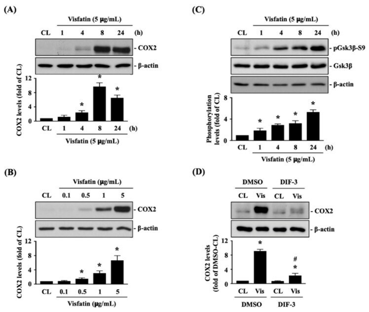
- Figure 1 Visfatin inactivates GSK3beta to increase catabolic COX2 expression in human primary chondrocytes. ( A - C ) Human primary chondrocytes were kept as controls (CL) or treated with 5 mug/mL of visfatin for 1, 4, 8 and 24 h or treated with 0.1, 0.5, 1 or 5 mug/mL of visfatin for 8 h and then ( A , B ) COX2 protein expression and ( C ) GSK3beta-Ser-9 phosphorylation were analyzed by Western blot. ( D ) Human primary chondrocytes were pretreated with DMSO or DIF-3, a GSK3beta activator, for 1 h and then kept as controls (CL) or treated with 5 mug/mL of visfatin (Vis) for 8 h. COX2 expression in human primary chondrocytes was analyzed by Western blot. Results in ( A - D ) are representative of three independent experiments with similar results. Statistical data in ( A - D ) are mean +- SEM from three independent experiments. *, p < 0.05 vs. untreated control cells (CL) or DMSO-CL-treated cells. #, p < 0.05 vs. DMSO-visfatin (Vis)-stimulated cells.
- Submitted by
- Invitrogen Antibodies (provider)
- Main image

- Experimental details
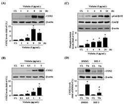
- Figure 2 p38 signaling regulates the effect of visfatin on GSK3beta inactivation and subsequent COX2 expression in human primary chondrocytes. ( A ) Human primary chondrocytes were kept as controls (CL) or treated with 5 mug/mL of visfatin for 1, 4, 8 and 24 h, and then the p38-Thr180/Tyr182 and ERK1/2-Thr202/Tyr204 kinases phosphorylations were analyzed by Western blot. ( B , C ) Human primary chondrocytes were pretreated with DMSO, PD98059 (ERK1/2 inhibitor, 30 muM), or SB203580 (p38 inhibitor, 10 muM) for 1 h and then kept as controls (CL) or treated with 5 mug/mL of visfatin (Vis) for 8 h. COX2 expression ( B ) and GSK3beta-Ser-9 phosphorylation ( C ) in human primary chondrocytes were analyzed by Western blot. Results in ( A - C ) are representative of three independent experiments with similar results. Statistic data in ( A - C ) are mean +- SEM from three independent experiments. *, p < 0.05 vs. untreated control cells (CL) or DMSO-CL-treated cells. #, p < 0.05 vs. DMSO-visfatin (Vis)-stimulated cells.

- Submitted by
- Invitrogen Antibodies (provider)
- Main image
- Experimental details
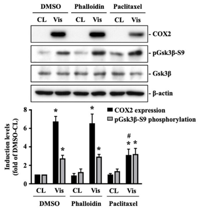
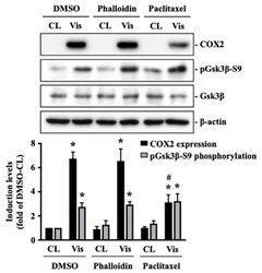
- Figure 5 Microtubule network damage influences the visfatin catabolic effect on COX2 upregulation in human primary chondrocytes. Human primary chondrocytes were pretreated with DMSO, phalloidin (microfilament stabilizer, 3 muM), or paclitaxel (microtubule stabilizer, 1 muM) for 1 h and then kept as controls (CL) or treated with 5 mug/mL of visfatin (Vis) for 8 h. COX2 expression and GSK3beta-Ser9 phosphorylation in human primary chondrocytes were analyzed by Western blot. Results are representative of three independent experiments with similar results. Statistic data are mean +- SEM from three independent experiments. *, p < 0.05 vs. DMSO-CL-treated cells. #, p < 0.05 vs. DMSO-visfatin (Vis)-stimulated cells.