Explore
 Validate
 Learn
Learn Immunocytochemistry
 Immunohistochemistry
Antibody data
- Antibody Data
- Antigen structure
- References [6]
- Comments [0]
- Validations
- Immunocytochemistry [1]
Submit
Validation data
Reference
Comment
Report error
- Product number
- HPA001335 - Provider product page

- Provider
- Atlas Antibodies
- Proper citation
- Atlas Antibodies Cat#HPA001335, RRID:AB_1079712
- Product name
- Anti-PTGS2
- Antibody type
- Polyclonal
- Description
- Polyclonal Antibody against Human PTGS2, Gene description: prostaglandin-endoperoxide synthase 2 (prostaglandin G/H synthase and cyclooxygenase), Alternative Gene Names: COX2, Validated applications: IHC, ICC, Uniprot ID: P35354, Storage: Store at +4°C for short term storage. Long time storage is recommended at -20°C.
- Reactivity
- Human
- Host
- Rabbit
- Conjugate
- Unconjugated
- Isotype
- IgG
- Vial size
- 100 µl
- Concentration
- 0.4 mg/ml
- Storage
- Store at +4°C for short term storage. Long time storage is recommended at -20°C.
- Handling
- The antibody solution should be gently mixed before use.
Submitted references Identification of AGR2 Gene-Specific Expression Patterns Associated with Epithelial-Mesenchymal Transition
Vasodilator oxyfedrine inhibits aldehyde metabolism and thereby sensitizes cancer cells to xCT-targeted therapy.
Understanding the Interplay between COX-2 and hTERT in Colorectal Cancer Using a Multi-Omics Analysis
Elevated levels of FN1 and CCL2 in bronchoalveolar lavage fluid from sarcoidosis patients
Prostaglandin E2 Regulates Pancreatic Stellate Cell Activity Via the EP4 Receptor
Wnt/β-Catenin Signaling Enhances Cyclooxygenase-2 (COX2) Transcriptional Activity in Gastric Cancer Cells
Martisova A, Sommerova L, Krejci A, Selingerova I, Kolarova T, Zavadil Kokas F, Holanek M, Podhorec J, Kazda T, Hrstka R
International Journal of Molecular Sciences 2022;23(18):10845
International Journal of Molecular Sciences 2022;23(18):10845
Vasodilator oxyfedrine inhibits aldehyde metabolism and thereby sensitizes cancer cells to xCT-targeted therapy.
Otsuki Y, Yamasaki J, Suina K, Okazaki S, Koike N, Saya H, Nagano O
Cancer science 2020 Jan;111(1):127-136
Cancer science 2020 Jan;111(1):127-136
Understanding the Interplay between COX-2 and hTERT in Colorectal Cancer Using a Multi-Omics Analysis
Ayiomamitis G, Notas G, Vasilakaki T, Tsavari A, Vederaki S, Theodosopoulos T, Kouroumalis E, Zaravinos A
Cancers 2019;11(10):1536
Cancers 2019;11(10):1536
Elevated levels of FN1 and CCL2 in bronchoalveolar lavage fluid from sarcoidosis patients
Hamsten C, Wiklundh E, Grönlund H, Schwenk J, Uhlén M, Eklund A, Nilsson P, Grunewald J, Häggmark-Månberg A
Respiratory Research 2016;17(1)
Respiratory Research 2016;17(1)
Prostaglandin E2 Regulates Pancreatic Stellate Cell Activity Via the EP4 Receptor
Charo C, Holla V, Arumugam T, Hwang R, Yang P, Dubois R, Menter D, Logsdon C, Ramachandran V
Pancreas 2013;42(3):467-474
Pancreas 2013;42(3):467-474
Wnt/β-Catenin Signaling Enhances Cyclooxygenase-2 (COX2) Transcriptional Activity in Gastric Cancer Cells
Campbell M, Nuñez F, Bravo S, Cruzat F, Montecino M, De Ferrari G
PLoS ONE 2011;6(4):e18562
PLoS ONE 2011;6(4):e18562
No comments: Submit comment
Supportive validation
- Submitted by
- Atlas Antibodies (provider)
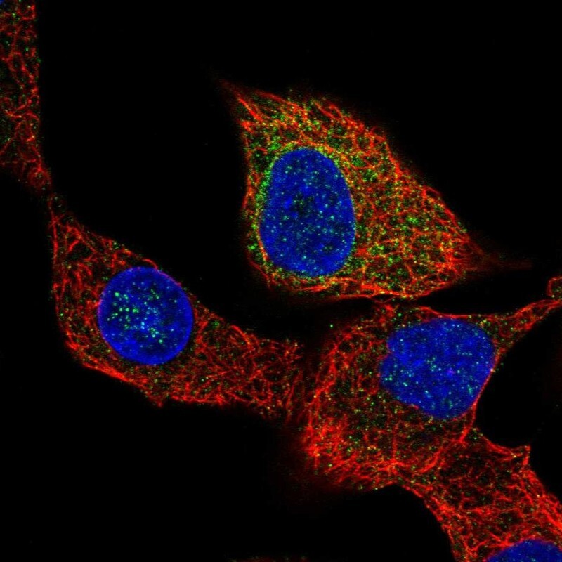
- Main image

- Experimental details
- Immunofluorescent staining of human cell line A549 shows localization to endoplasmic reticulum.
- Sample type
- Human