Explore
 Validate
 Learn
Learn Western blot
Antibody data
- Antibody Data
- Antigen structure
- References [0]
- Comments [0]
- Validations
- Western blot [6]
- Immunocytochemistry [1]
- Immunohistochemistry [1]
Submit
Validation data
Reference
Comment
Report error
- Product number
- PA5-27238 - Provider product page

- Provider
- Invitrogen Antibodies
- Product name
- COX2 Polyclonal Antibody
- Antibody type
- Polyclonal
- Antigen
- Synthetic peptide
- Description
- Recommended positive controls: THP-1 (100 ng/mL PMA treatment for 16 hr and 1000 ng/mL LPS treatment for 24 hr), RAW264.7 (0.1 µg/mL PMA treatment for 16 hr and 1 µg/mL LPS treatment for 24 hr).
- Concentration
- 0.42 mg/mL
No comments: Submit comment
Supportive validation
- Submitted by
- Invitrogen Antibodies (provider)
- Main image
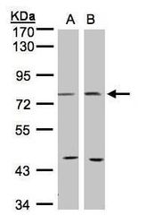
- Experimental details
- Western blot analysis of COX2 using 30 µg of A) 293T and B) MOLT4 lysate. Samples were loaded onto a 7.5% SDS-PAGE gel and probed with a COX2 polyclonal antibody (Product # PA5-27238) at a dilution of 1:1000.

- Submitted by
- Invitrogen Antibodies (provider)
- Main image
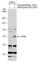
- Experimental details
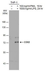
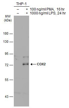
- Western Blot using COX2 Polyclonal Antibody (Product # PA5-27238). Untreated (–) and treated (+) THP-1 whole cell extracts (30 µg) were separated by 7.5% SDS-PAGE, and the membrane was blotted with COX2 Polyclonal Antibody (Product # PA5-27238) diluted at 1:500. The HRP-conjugated anti-rabbit IgG antibody was used to detect the primary antibody.
- Submitted by
- Invitrogen Antibodies (provider)
- Main image
- Experimental details
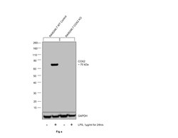
- Knockout of COX2 was achieved by CRISPR-Cas9 genome editing using TrueGuide Synthetic gRNA (Product # A35533, Assay ID CRISPR568234_SGM) and LentiArray Cas9 Lentivirus (Product # A32064). Western blot analysis of COX2 was performed by loading 30 µg of RAW 246.7 wild type (Lane 1), RAW 246.7 wild type cells treated with 1 µg/mL LPS for 24hrs (Lane 2), RAW 246.7 COX2 KO (Lane 3) and RAW 246.7 COX2 KO treated with 1 µg/mL LPS for 24hrs (Lane 4) membrane enriched extracts. The samples were electrophoresed using NuPAGE™ Novex™ 4-12% Bis-Tris Protein Gel (Product # NP0322BOX). Resolved proteins were then transferred onto a nitrocellulose membrane (Product # IB23001) by iBlot® 2 Dry Blotting System (Product # IB21001). The blot was probed with COX2 Polyclonal Antibody (Product # PA5-27238, 1:3000 dilution) and Goat anti-Rabbit IgG (H+L) Superclonal™ Recombinant Secondary Antibody, HRP (Product # A27036, 1:10,000 dilution) using the iBright™ FL 1500 (Product # A44115). Chemiluminescent detection was performed using SuperSignal™ West Dura Extended Duration Substrate (Product # 34076). Loss of signal upon CRISPR mediated knockout (KO) using the LentiArray™ CRISPR product line confirms that antibody is specific to COX2.

- Submitted by
- Invitrogen Antibodies (provider)
- Main image
- Experimental details
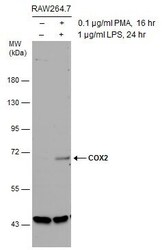
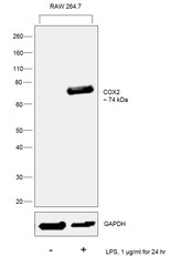
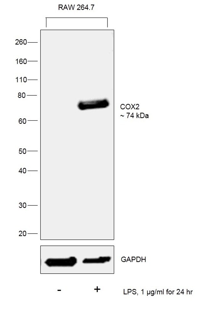
- Western Blot analysis of COX2 was performed by separating 30 µg of untreated (–) and treated (+) RAW264.7 whole cell extracts by 5% SDS-PAGE. Proteins were transferred to a membrane and probed with a COX2 Polyclonal Antibody (Product # PA5-27238) at a dilution of 1:1000.
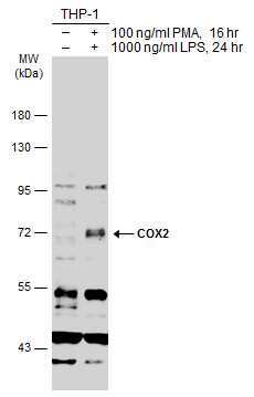
- Submitted by
- Invitrogen Antibodies (provider)
- Main image
- Experimental details
- Western Blot using COX2 Polyclonal Antibody (Product # PA5-27238). Untreated (–) and treated (+) THP-1 whole cell extracts (30 µg) were separated by 7.5% SDS-PAGE, and the membrane was blotted with COX2 Polyclonal Antibody (Product # PA5-27238) diluted at 1:500. The HRP-conjugated anti-rabbit IgG antibody was used to detect the primary antibody.
- Submitted by
- Invitrogen Antibodies (provider)
- Main image
- Experimental details
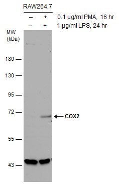
- Western blot was performed using Anti-COX2 Rabbit Polyclonal Antibody (Product # PA5-27238) and a 74 kDa band corresponding to COX2 was induced in RAW 264.7 upon LPS treatment. Membrane enriched extracts (30 µg lysate) of RAW 264.7 (Lane 1) and RAW 264.7 treated with LPS (1µg/ml for 24 hr) (Lane 2) were electrophoresed using Novex® NuPAGE® 12 % Bis-Tris gel (Product # NP0342BOX). Resolved proteins were then transferred onto a nitrocellulose membrane (Product # IB23001) by iBlot® 2 Dry Blotting System (Product # IB21001). The blot was probed with the primary antibody (1:3000 dilution) and detected by chemiluminescence with Goat anti-Rabbit IgG (H+L), Superclonal™ Recombinant Secondary Antibody, HRP (Product # A27036, 1:4000 dilution) using the iBright FL 1000 (Product # A32752). Chemiluminescent detection was performed using Novex® ECL Chemiluminescent Substrate Reagent Kit (Product # WP20005)..
Supportive validation
- Submitted by
- Invitrogen Antibodies (provider)
- Main image
- Experimental details
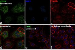
- Immunofluorescence analysis of COX2 was performed using 70% confluent log phase RAW 264.7 cells treated with 100 ng/mL of Lipopolysaccharide for 24 Hrs. The cells were fixed with 4% Paraformaldehyde for 10 minutes, permeabilized with 0.1% Triton™ X-100 for 10 minutes, and blocked with 2% BSA for 10 minutes at room temperature. The cells were labeled with COX2 Polyclonal Antibody (Product # PA5-27238) at 10 µg/mL in 0.1% BSA, incubated at 4 degree celsius overnight and then labeled with Goat anti-Rabbit IgG (H+L), Superclonal™ Recombinant Secondary Antibody, Alexa Fluor 488 (Product # A27034), (1:2000 dilution) for 45 minutes at room temperature (Panel a: Green). Nuclei (Panel b: Blue) were stained with SlowFade® Gold Antifade Mountant with DAPI (Product # S36938). F-actin (Panel c: Red) was stained with Rhodamine Phalloidin (Product # R415, 1:300). Panel d represents the merged image showing Cytoplasmic and nuclear membrane localization. Panel e represents untreated cells with reduced signal. Panel f represents control cells with no primary antibody to assess background. The images were captured at 60X magnification.

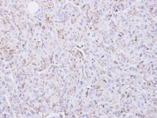
Supportive validation
- Submitted by
- Invitrogen Antibodies (provider)
- Main image
- Experimental details
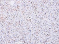
- Immunohistochemical analysis of paraffin-embedded U87 xenograft, using COX2 (Product # PA5-27238) antibody at 1:100 dilution. Antigen Retrieval: Citrate buffer, pH 6.0, 15 min.