Explore
 Validate
 Learn
Learn Western blot
 ELISA
ELISAAntibody data
- Antibody Data
- Antigen structure
- References [4]
- Comments [0]
- Validations
- Western blot [1]
- Immunocytochemistry [1]
- Immunohistochemistry [2]
Submit
Validation data
Reference
Comment
Report error
- Product number
- 11836-1-AP - Provider product page

- Provider
- Proteintech Group
- Proper citation
- Proteintech Cat#11836-1-AP, RRID:AB_2097479
- Product name
- LBP antibody
- Antibody type
- Polyclonal
- Description
- LBP antibody (Cat. #11836-1-AP) is a rabbit polyclonal antibody that shows reactivity with human, mouse, rat and has been validated for the following applications: FC, IF, IHC, IP, WB,ELISA.
- Reactivity
- Human, Mouse, Rat
- Host
- Rabbit
- Conjugate
- Unconjugated
- Isotype
- IgG
- Vial size
- 20ul, 150ul
Submitted references Dysregulation of FLVCR1a-dependent mitochondrial calcium handling in neural progenitors causes congenital hydrocephalus.
The characteristic regulation of gene expression Lbp and Sod3 in peri-implant connective tissue of rats.
Association of decreased levels of lipopolysaccharide-binding protein with OKN-007-induced regression of tumor growth in an F98 rat glioma model.
Circulating exosomes contain protein biomarkers of metastatic non-small-cell lung cancer.
Bertino F, Mukherjee D, Bonora M, Bagowski C, Nardelli J, Metani L, Zanin Venturini DI, Chianese D, Santander N, Salaroglio IC, Hentschel A, Quarta E, Genova T, McKinney AA, Allocco AL, Fiorito V, Petrillo S, Ammirata G, De Giorgio F, Dennis E, Allington G, Maier F, Shoukier M, Gloning KP, Munaron L, Mussano F, Salsano E, Pareyson D, di Rocco M, Altruda F, Panagiotakos G, Kahle KT, Gressens P, Riganti C, Pinton PP, Roos A, Arnold T, Tolosano E, Chiabrando D
Cell reports. Medicine 2024 Jul 16;5(7):101647
Cell reports. Medicine 2024 Jul 16;5(7):101647
The characteristic regulation of gene expression Lbp and Sod3 in peri-implant connective tissue of rats.
Kobayashi T, Sasaki H, Asami Y, Mori G, Yoshinari M, Yajima Y
Journal of biomedical materials research. Part A 2020 Mar;108(3):592-600
Journal of biomedical materials research. Part A 2020 Mar;108(3):592-600
Association of decreased levels of lipopolysaccharide-binding protein with OKN-007-induced regression of tumor growth in an F98 rat glioma model.
Smith N, Saunders D, Jensen RL, Towner RA
Journal of neurosurgery 2020 Dec 1;133(6):1695-1703
Journal of neurosurgery 2020 Dec 1;133(6):1695-1703
Circulating exosomes contain protein biomarkers of metastatic non-small-cell lung cancer.
Wang N, Song X, Liu L, Niu L, Wang X, Song X, Xie L
Cancer science 2018 May;109(5):1701-1709
Cancer science 2018 May;109(5):1701-1709
No comments: Submit comment
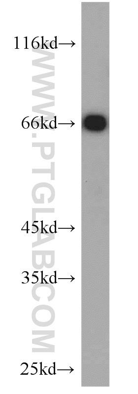
Supportive validation
- Submitted by
- Proteintech Group (provider)
- Main image

- Experimental details
- mouse liver tissue were subjected to SDS PAGE followed by western blot with 11836-1-AP(LBP antibody) at dilution of 1:1000
- Sample type
- tissue
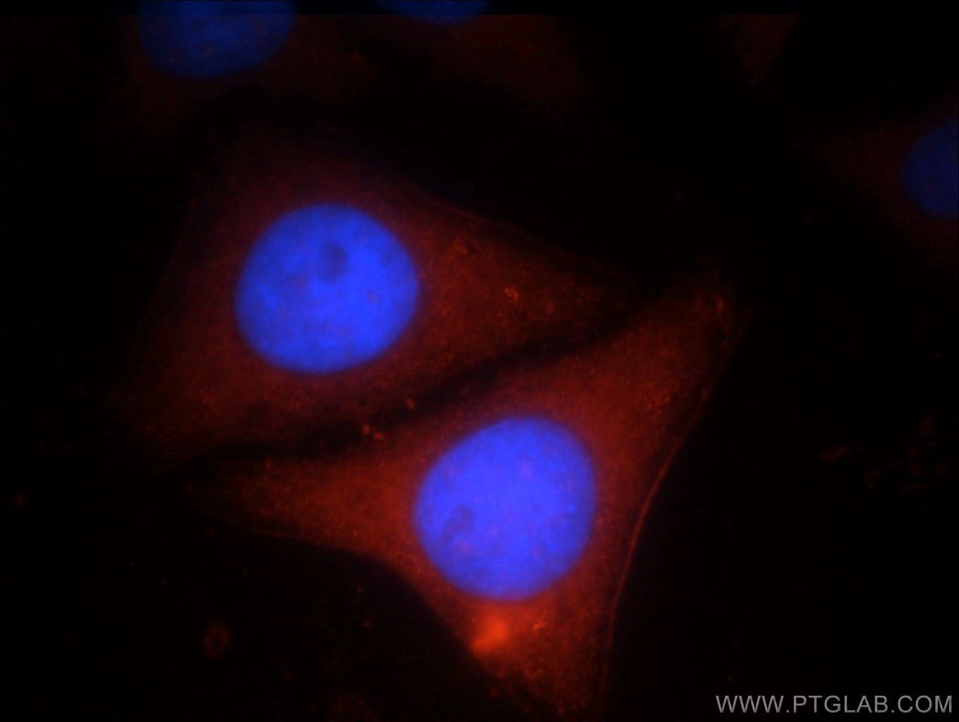
Supportive validation
- Submitted by
- Proteintech Group (provider)
- Main image

- Experimental details
- Immunofluorescent analysis of HepG2 cells, using LBP antibody 11836-1-AP at 1:25 dilution and Rhodamine-labeled goat anti-rabbit IgG (red). Blue pseudocolor = DAPI (fluorescent DNA dye).
- Sample type
- cell line
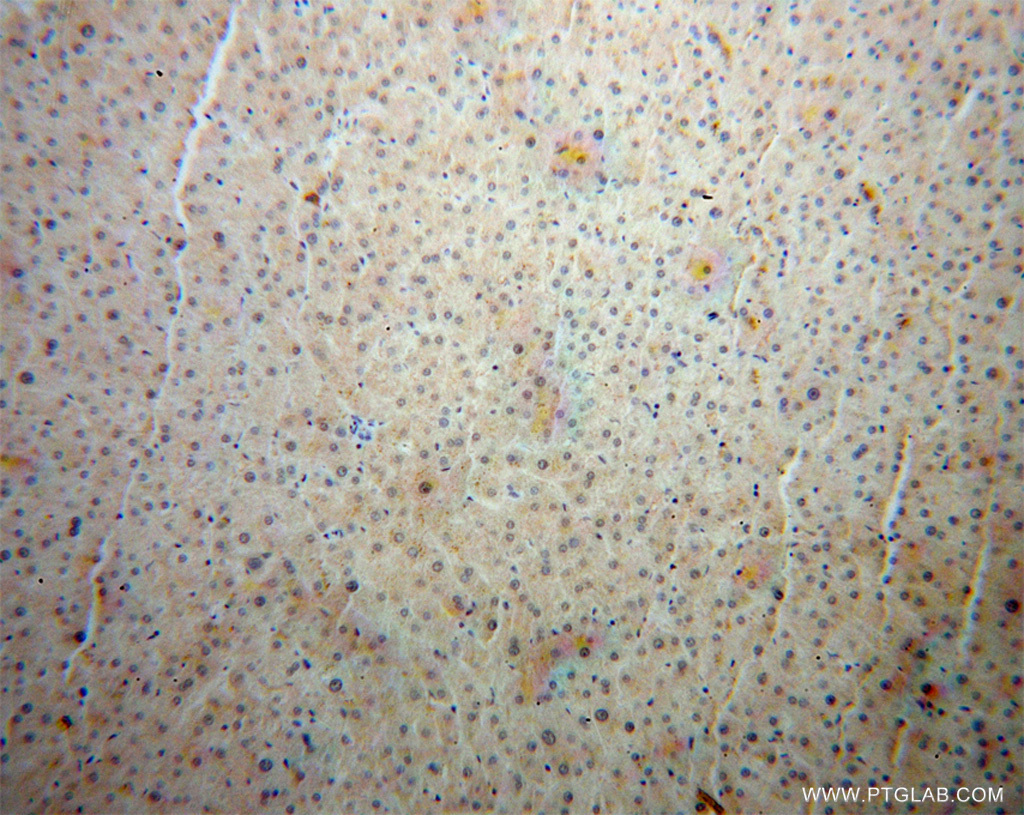
Supportive validation
- Submitted by
- Proteintech Group (provider)
- Main image

- Experimental details
- Immunohistochemical of paraffin-embedded human liver cancer using 11836-1-AP(LBP antibody) at dilution of 1:50 (under 10x lens)
- Sample type
- tissue
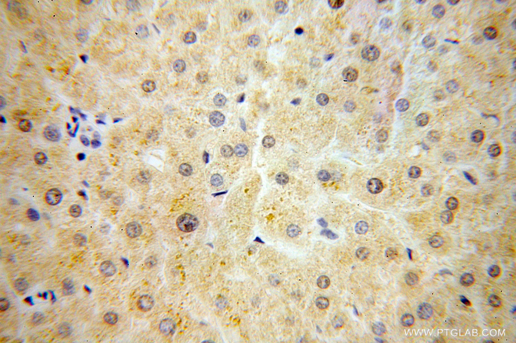
- Submitted by
- Proteintech Group (provider)
- Main image
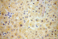
- Experimental details
- Immunohistochemical of paraffin-embedded human liver cancer using 11836-1-AP(LBP antibody) at dilution of 1:50 (under 40x lens)
- Sample type
- tissue