 Explore
 Validate
 Learn
Learn Flow cytometry
Antibody data
- Antibody Data
- Antigen structure
- References [2]
- Comments [0]
- Validations
- Flow cytometry [2]
Submit
Validation data
Reference
Comment
Report error
- Product number
- IC8026P - Provider product page

- Provider
- R&D Systems
- Product name
- Human Arginase 1/ARG1 PE-conjugated Antibody
- Antibody type
- Monoclonal
- Description
- Protein A or G purified from hybridoma culture supernatant. Detects human Arginase 1/ARG1 in ELISAs.
- Reactivity
- Human
- Host
- Mouse
- Antigen sequence
P05089- Isotype
- IgG
- Antibody clone number
- 658922
- Vial size
- 100 Tests
- Storage
- Protect from light. Do not freeze. 12 months from date of receipt, 2 to 8 °C as supplied.
Submitted references Monocyte Differentiation towards Protumor Activity Does Not Correlate with M1 or M2 Phenotypes.
Ubiquitous Over-Expression of Chromatin Remodeling Factor SRG3 Ameliorates the T Cell-Mediated Exacerbation of EAE by Modulating the Phenotypes of both Dendritic Cells and Macrophages.
Chimal-Ramírez GK, Espinoza-Sánchez NA, Chávez-Sánchez L, Arriaga-Pizano L, Fuentes-Pananá EM
Journal of immunology research 2016;2016:6031486
Journal of immunology research 2016;2016:6031486
Ubiquitous Over-Expression of Chromatin Remodeling Factor SRG3 Ameliorates the T Cell-Mediated Exacerbation of EAE by Modulating the Phenotypes of both Dendritic Cells and Macrophages.
Lee SW, Park HJ, Jeon SH, Lee C, Seong RH, Park SH, Hong S
PloS one 2015;10(7):e0132329
PloS one 2015;10(7):e0132329
No comments: Submit comment
Supportive validation
- Submitted by
- R&D Systems (provider)
- Main image
- Experimental details
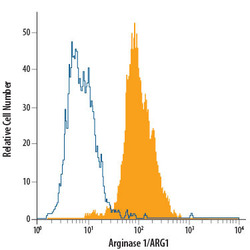
- Detection of Arginase 1/ARG1 in HepG2 Human Cell Line by Flow Cytometry. HepG2 human hepatocellular carcinoma cell line was stained with Mouse Anti-Human Arginase 1/ARG1 PE-conjugated Mono-clonal Antibody (Catalog # IC8026P, filled histogram) or isotype control antibody (Catalog # IC0041P, open histogram). To facilitate intracellular staining, cells were fixed with Flow Cytometry Fixation Buffer (Catalog # FC004) and permeabilized with Flow Cytometry Permeabilization/Wash Buffer I (Catalog # FC005). View our protocol for Staining Intracellular Molecules.

- Submitted by
- R&D Systems (provider)
- Main image

- Experimental details
- Detection of Arginase 1/ARG1 in Human PBMCs by Flow Cytometry. Human peripheral blood mononuclear cells (PBMCs) were stained with Mouse Anti-Human Integrin alpha M/CD11b APC-conjugated Monoclonal Antibody (Catalog # FAB1699A) and either (A) Mouse Anti-Human Arginase 1/ARG1 PE-conjugated Monoclonal Antibody (Catalog # IC8026P) or (B) Mouse IgG2B Phycoerythrin Isotype Control (Catalog # IC0041P). To facilitate intracellular staining, cells were fixed with Flow Cytometry Fixation Buffer (Catalog # FC004) and permeabilized with Flow Cytometry Permeabilization/Wash Buffer I (Catalog # FC005). View our protocol for Staining Intracellular Molecules.
