 Explore
 Validate
 Learn
Learn Western blot
 ELISA
ELISAAntibody data
- Antibody Data
- Antigen structure
- References [9]
- Comments [0]
- Validations
- Western blot [4]
Submit
Validation data
Reference
Comment
Report error
- Product number
- NB100-59740 - Provider product page

- Provider
- Novus Biologicals
- Proper citation
- Novus Cat#NB100-59740, RRID:AB_2058951
- Product name
- Goat Polyclonal Arginase 1/ARG1/liver Arginase Antibody
- Antibody type
- Polyclonal
- Description
- Immunogen affinity purified.
- Reactivity
- Human, Mouse, Rat
- Host
- Goat
- Antigen sequence
NHKPETDYLKPPK- Isotype
- IgG
- Vial size
- 0.1 mg
- Concentration
- 0.5 mg/ml
- Storage
- Store at -20C. Avoid freeze-thaw cycles.
Submitted references Short-Chain Fatty Acids Improve Poststroke Recovery via Immunological Mechanisms.
PC1/3 KD Macrophages Exhibit Resistance to the Inhibitory Effect of IL-10 and a Higher TLR4 Activation Rate, Leading to an Anti-Tumoral Phenotype.
Metabolic origins of spatial organization in the tumor microenvironment.
Phenotypic polarization of activated astrocytes: the critical role of lipocalin-2 in the classical inflammatory activation of astrocytes.
Secreted protein lipocalin-2 promotes microglial M1 polarization.
All-trans retinoic acid modifies the expression of clock and disease marker genes.
Caffeine alters circadian rhythms and expression of disease and metabolic markers.
Identification of an IFN-γ/mast cell axis in a mouse model of chronic asthma.
Arginase-flotillin interaction brings arginase to red blood cell membrane.
Sadler R, Cramer JV, Heindl S, Kostidis S, Betz D, Zuurbier KR, Northoff BH, Heijink M, Goldberg MP, Plautz EJ, Roth S, Malik R, Dichgans M, Holdt LM, Benakis C, Giera M, Stowe AM, Liesz A
The Journal of neuroscience : the official journal of the Society for Neuroscience 2020 Jan 29;40(5):1162-1173
The Journal of neuroscience : the official journal of the Society for Neuroscience 2020 Jan 29;40(5):1162-1173
PC1/3 KD Macrophages Exhibit Resistance to the Inhibitory Effect of IL-10 and a Higher TLR4 Activation Rate, Leading to an Anti-Tumoral Phenotype.
Rodet F, Capuz A, Ozcan BA, Le Beillan R, Raffo-Romero A, Kobeissy F, Duhamel M, Salzet M
Cells 2019 Nov 22;8(12)
Cells 2019 Nov 22;8(12)
Metabolic origins of spatial organization in the tumor microenvironment.
Carmona-Fontaine C, Deforet M, Akkari L, Thompson CB, Joyce JA, Xavier JB
Proceedings of the National Academy of Sciences of the United States of America 2017 Mar 14;114(11):2934-2939
Proceedings of the National Academy of Sciences of the United States of America 2017 Mar 14;114(11):2934-2939
Phenotypic polarization of activated astrocytes: the critical role of lipocalin-2 in the classical inflammatory activation of astrocytes.
Jang E, Kim JH, Lee S, Kim JH, Seo JW, Jin M, Lee MG, Jang IS, Lee WH, Suk K
Journal of immunology (Baltimore, Md. : 1950) 2013 Nov 15;191(10):5204-19
Journal of immunology (Baltimore, Md. : 1950) 2013 Nov 15;191(10):5204-19
Secreted protein lipocalin-2 promotes microglial M1 polarization.
Jang E, Lee S, Kim JH, Kim JH, Seo JW, Lee WH, Mori K, Nakao K, Suk K
FASEB journal : official publication of the Federation of American Societies for Experimental Biology 2013 Mar;27(3):1176-90
FASEB journal : official publication of the Federation of American Societies for Experimental Biology 2013 Mar;27(3):1176-90
All-trans retinoic acid modifies the expression of clock and disease marker genes.
Sherman H, Gutman R, Chapnik N, Meylan J, le Coutre J, Froy O
The Journal of nutritional biochemistry 2012 Mar;23(3):209-17
The Journal of nutritional biochemistry 2012 Mar;23(3):209-17
Caffeine alters circadian rhythms and expression of disease and metabolic markers.
Sherman H, Gutman R, Chapnik N, Meylan J, le Coutre J, Froy O
The international journal of biochemistry & cell biology 2011 May;43(5):829-38
The international journal of biochemistry & cell biology 2011 May;43(5):829-38
Identification of an IFN-γ/mast cell axis in a mouse model of chronic asthma.
Yu M, Eckart MR, Morgan AA, Mukai K, Butte AJ, Tsai M, Galli SJ
The Journal of clinical investigation 2011 Aug;121(8):3133-43
The Journal of clinical investigation 2011 Aug;121(8):3133-43
Arginase-flotillin interaction brings arginase to red blood cell membrane.
Jiang M, Ding Y, Su Y, Hu X, Li J, Zhang Z
FEBS letters 2006 Dec 11;580(28-29):6561-4
FEBS letters 2006 Dec 11;580(28-29):6561-4
No comments: Submit comment
Supportive validation
- Submitted by
- Novus Biologicals (provider)
- Main image
- Experimental details
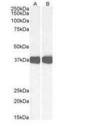
- Western Blot: Arginase 1/ARG1/liver Arginase Antibody [NB100-59740] - Staining of Mouse Liver (A) and Rat Liver (B) lysates (35 ug protein in RIPA buffer). Primary incubation was 1 hour. Detected by chemiluminescence.

- Submitted by
- Novus Biologicals (provider)
- Main image
- Experimental details
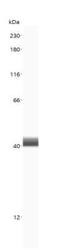
- Simple Western: Arginase 1/ARG1/liver Arginase Antibody [NB100-59740] - Simple Western lane view shows a specific band for ARG1 in mouse Liver lysate using ARG1 antibody (NB100-59740) at 5 ug/mL. This experiment was performed under reducing conditions using the 12-230 kDa separation system.

- Submitted by
- Novus Biologicals (provider)
- Main image
- Experimental details
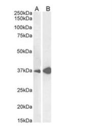
- Western Blot: Arginase 1/ARG1/liver Arginase Antibody [NB100-59740] - Staining of Mouse Liver (A) and Rat Liver (B) lysates (35ug protein in RIPA buffer). Detected by chemiluminescence.

- Submitted by
- Novus Biologicals (provider)
- Main image
- Experimental details
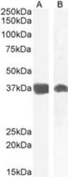
- Western Blot: Arginase 1/ARG1/liver Arginase Antibody [NB100-59740] - Staining of Mouse (A) and Rat (B) Liver lysate (35 ug protein in RIPA buffer). Antibody at 0.01 ug/mL. Detected by chemiluminescence.
