Explore
 Validate
 Learn
Learn Western blot
Antibody data
- Antibody Data
- Antigen structure
- References [7]
- Comments [0]
- Validations
- Western blot [2]
- Immunohistochemistry [1]
- Other assay [1]
Submit
Validation data
Reference
Comment
Report error
- Product number
- 14-9779-82 - Provider product page

- Provider
- Invitrogen Antibodies
- Product name
- Arginase 1 Monoclonal Antibody (sl6arg), eBioscience™
- Antibody type
- Monoclonal
- Antigen
- Other
- Description
- Description: The monoclonal antibody sl6arg recognizes human Arginase-1, a 35 kDa enzyme that that helps eliminate nitrogen by converting L-arginine to urea and L-ornithine which further converts to polyamine. These polyamines are important for cell proliferation and removal of toxins that arise from protein degradation. Expression is found in the liver, neutrophils, as well as myeloid derived suppressor cells (MDSC) and neural stem cells. Expression in macrophages has not been determined in humans, while in mice expression in M2 macrophages is well established. Arginase-1 may be expressed in the myeloid population in breast cancer tumors and is typically found in the majority of hepatocellular carcinomas.
- Antibody clone number
- sl6arg
- Concentration
- 0.5 mg/mL
Submitted references Pulmonary Conventional Type 1 Langerin-Expressing Dendritic Cells Play a Role in Impairing Early Protective Immune Response against Cryptococcus neoformans Infection in Mice.
Deficient Adipogenesis of Scleroderma Patient and Healthy African American Monocytes.
Metabolism of L-arginine by myeloid-derived suppressor cells in cancer: mechanisms of T cell suppression and therapeutic perspectives.
Arginase-1 is a more sensitive marker of hepatic differentiation than HepPar-1 and glypican-3 in fine-needle aspiration biopsies.
Characterization of cytokine-induced myeloid-derived suppressor cells from normal human peripheral blood mononuclear cells.
Arginase-1-expressing macrophages suppress Th2 cytokine-driven inflammation and fibrosis.
Alternative metabolic states in murine macrophages reflected by the nitric oxide synthase/arginase balance: competitive regulation by CD4+ T cells correlates with Th1/Th2 phenotype.
Guasconi L, Beccacece I, Volpini X, Burstein VL, Mena CJ, Silvane L, Almeida MA, Musri MM, Cervi L, Chiapello LS
Journal of fungi (Basel, Switzerland) 2022 Jul 28;8(8)
Journal of fungi (Basel, Switzerland) 2022 Jul 28;8(8)
Deficient Adipogenesis of Scleroderma Patient and Healthy African American Monocytes.
Lee R, Reese C, Carmen-Lopez G, Perry B, Bonner M, Zemskova M, Wilson CL, Helke KL, Silver RM, Hoffman S, Tourkina E
Frontiers in pharmacology 2017;8:174
Frontiers in pharmacology 2017;8:174
Metabolism of L-arginine by myeloid-derived suppressor cells in cancer: mechanisms of T cell suppression and therapeutic perspectives.
Raber P, Ochoa AC, Rodríguez PC
Immunological investigations 2012;41(6-7):614-34
Immunological investigations 2012;41(6-7):614-34
Arginase-1 is a more sensitive marker of hepatic differentiation than HepPar-1 and glypican-3 in fine-needle aspiration biopsies.
Fujiwara M, Kwok S, Yano H, Pai RK
Cancer cytopathology 2012 Aug 25;120(4):230-7
Cancer cytopathology 2012 Aug 25;120(4):230-7
Characterization of cytokine-induced myeloid-derived suppressor cells from normal human peripheral blood mononuclear cells.
Lechner MG, Liebertz DJ, Epstein AL
Journal of immunology (Baltimore, Md. : 1950) 2010 Aug 15;185(4):2273-84
Journal of immunology (Baltimore, Md. : 1950) 2010 Aug 15;185(4):2273-84
Arginase-1-expressing macrophages suppress Th2 cytokine-driven inflammation and fibrosis.
Pesce JT, Ramalingam TR, Mentink-Kane MM, Wilson MS, El Kasmi KC, Smith AM, Thompson RW, Cheever AW, Murray PJ, Wynn TA
PLoS pathogens 2009 Apr;5(4):e1000371
PLoS pathogens 2009 Apr;5(4):e1000371
Alternative metabolic states in murine macrophages reflected by the nitric oxide synthase/arginase balance: competitive regulation by CD4+ T cells correlates with Th1/Th2 phenotype.
Munder M, Eichmann K, Modolell M
Journal of immunology (Baltimore, Md. : 1950) 1998 Jun 1;160(11):5347-54
Journal of immunology (Baltimore, Md. : 1950) 1998 Jun 1;160(11):5347-54
No comments: Submit comment
Supportive validation
- Submitted by
- Invitrogen Antibodies (provider)
- Main image
- Experimental details
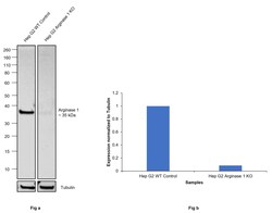
- Knockout of Arginase 1 was achieved by CRISPR-Cas9 genome editing using LentiArray™ Lentiviral sgRNA (Product # A32042, Assay ID CRIPSR760199_LV) and LentiArray Cas9 Lentivirus (Product # A32064). Western blot analysis of Arginase 1 was performed by loading 30 µg of Hep G2 wild type (Lane 1) and Hep G2 Arginase 1 KO (Lane 3) whole cell extracts. The samples were electrophoresed using NuPAGE™ Novex™ 4-12% Bis-Tris Protein Gel (Product # NP0321BOX). Resolved proteins were then transferred onto a nitrocellulose membrane (Product # IB23001) by iBlot® 2 Dry Blotting System (Product # IB21001). The blot was probed with Arginase 1 Monoclonal Antibody (sl6arg), eBioscience™ (Product # 14-9779-82, 1:500 dilution) and Goat anti-Mouse IgG (H+L) Superclonal™ Recombinant Secondary Antibody, HRP (Product # A28177, 1:10,000 dilution). Chemiluminescent detection was performed using SuperSignal™ West Atto Ultimate Sensitivity Substrate (Product # A38556) with the iBright™ FL1500 (Product # A44115). Loss of signal upon CRISPR mediated knockout (KO) using the LentiArray™ CRISPR product line confirms that antibody is specific to Arginase 1.

- Submitted by
- Invitrogen Antibodies (provider)
- Main image

- Experimental details
- Western blot was performed using Anti-Arginase 1 Monoclonal Antibody (sl6arg), eBioscience™ (Product # 14-9779-82) and a 35kDa band corresponding to Arginase-1 was observed. Whole cell extracts (40 µg lysate) of MOLT-4 (Lane 1) and Hep G2 (Lane 2) were electrophoresed using NuPAGE™ 4-12% Bis-Tris Protein Gel (Product # NP0322BOX). Resolved proteins were then transferred onto a Nitrocellulose membrane (Product # IB23001) by iBlot® 2 Dry Blotting System (Product # IB21001). The blot was probed with the primary antibody (1:500) and detected by chemiluminescence with Goat anti-Mouse IgG (H+L) Superclonal™ Recombinant Secondary Antibody, HRP (Product # A28177, 1:4000) using the iBright FL 1000 (Product # A32752). Chemiluminescent detection was performed using SuperSignal™ West Dura Extended Duration Substrate (Product # 34076).

Supportive validation
- Submitted by
- Invitrogen Antibodies (provider)
- Main image
- Experimental details
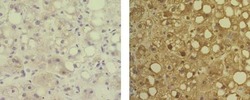
- Immunohistochemistry of formalin-fixed paraffin embedded human liver tissue using 5 µg/mL of Mouse IgG1 K Isotype Control Purified (Product # 14-4714-82) (left) or 5 µg/mL of Anti-Human Arginase-1 Purified (right), followed by Anti-Mouse IgG Biotin, Streptavidin HRP, and DAB visualization.Nuclei are counterstained with hematoxylin.

Supportive validation
- Submitted by
- Invitrogen Antibodies (provider)
- Main image
- Experimental details
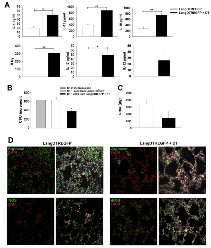
- Ex vivo cytokine production, fungal growth inhibition and arginase-1 or iNOS expression by lung cells from C. neoformans -infected mice. ( A ) Cytokine production by lung cell suspensions obtained from DT-treated (black bars) or untreated (white bars) LangDTREGFP mice at 7 days post infection. Bars represent cytokine levels (ELISA) in 24 h culture supernatants. * p < 0.05; ** p < 0.005. ( B ) In vitro C. neoformans growth assay using a yeast suspension (1 x 10 3 ) cultured in medium alone (gray bar) or in the presence of adherent lung cells from untreated (white bar) or DT-treated (black bar) 7-day-infected LangDTREGFP mice. Bars represent ratio of final vs initial CFU numbers in each culture condition. ( C ) Arginase activity from lysates of adherent cells cultured in ( B ), measured as urea production (ug of urea per ug of protein). * p < 0.05. Data are expressed as mean +- SEM. The data shown are pooled from two independent experiments ( n = 6 animals per group; samples from each animal were analyzed in triplicate ( A , C ) or duplicate ( B )). All data were analyzed with Student's t -test or ANOVA. ( D ) Confocal microscopy of representative lung sections from 7-day-infected mice ( n = 6) after immunostaining with fluorochrome-labeled specific antibodies to arginase-1 (upper panels) or iNOS (lower panels). 600x magnification. DT: diphtheria toxin; dpi: days post infection; CFU: colony-forming units.