Explore
 Validate
 Learn
Learn Western blot
 Immunocytochemistry
Antibody data
- Antibody Data
- Antigen structure
- References [1]
- Comments [0]
- Validations
- Immunocytochemistry [1]
- Other assay [1]
Submit
Validation data
Reference
Comment
Report error
- Product number
- 53-9779-82 - Provider product page

- Provider
- Invitrogen Antibodies
- Product name
- Arginase 1 Monoclonal Antibody (sl6arg), Alexa Fluor™ 488, eBioscience™
- Antibody type
- Monoclonal
- Antigen
- Other
- Description
- Description: The monoclonal antibody sl6arg recognizes human Arginase-1, a 35 kDa enzyme that that helps eliminate nitrogen by converting L-arginine to urea and L-ornithine which further converts to polyamine. These polyamines are important for cell proliferation and removal of toxins that arise from protein degradation. Expression is found in the liver, neutrophils, as well as myeloid derived suppressor cells (MDSC) and neural stem cells. Expression in macrophages has not been determined in humans, while in mice expression in M2 macrophages is well established. Arginase-1 may be expressed in the myeloid population in breast cancer tumors and is typically found in the majority of hepatocellular carcinomas. Applications Reported: This sl6arg antibody has been reported for use in immunohistochemical staining of formalin-fixed paraffin embedded tissue sections, microscopy, and immunocytochemistry. Applications Tested: This sl6arg antibody has been tested by immunocytochemistry of formaldehyde-fixed and permeabilized cells and can be used at less than or equal to 20 µg/mL. It is recommended that the antibody be carefully titrated for optimal performance in the assay of interest. Excitation: 488 nm; Emission: 519 nm; Laser: Blue Laser. Filtration: 0.2 µm post-manufacturing filtered.
- Reactivity
- Human
- Host
- Mouse
- Conjugate
- Green dye
- Isotype
- IgG
- Antibody clone number
- sl6arg
- Vial size
- 100 µg
- Concentration
- 0.5 mg/mL
- Storage
- 4° C, store in dark, DO NOT FREEZE!
Submitted references Pulmonary Conventional Type 1 Langerin-Expressing Dendritic Cells Play a Role in Impairing Early Protective Immune Response against Cryptococcus neoformans Infection in Mice.
Guasconi L, Beccacece I, Volpini X, Burstein VL, Mena CJ, Silvane L, Almeida MA, Musri MM, Cervi L, Chiapello LS
Journal of fungi (Basel, Switzerland) 2022 Jul 28;8(8)
Journal of fungi (Basel, Switzerland) 2022 Jul 28;8(8)
No comments: Submit comment
Supportive validation
- Submitted by
- Invitrogen Antibodies (provider)
- Main image

- Experimental details
- Immunocytochemistry of fixed and permeabilized HepG2 cells stained with 20 µg/mL Mouse IgG1 K Isotype Control Alexa Fluor® 488 (left) or 20 µg/mL Anti-Human Arginase-1 Alexa Fluor® 488 (right). Nuclei are stained with DAPI.
- Conjugate
- Green dye

Supportive validation
- Submitted by
- Invitrogen Antibodies (provider)
- Main image
- Experimental details
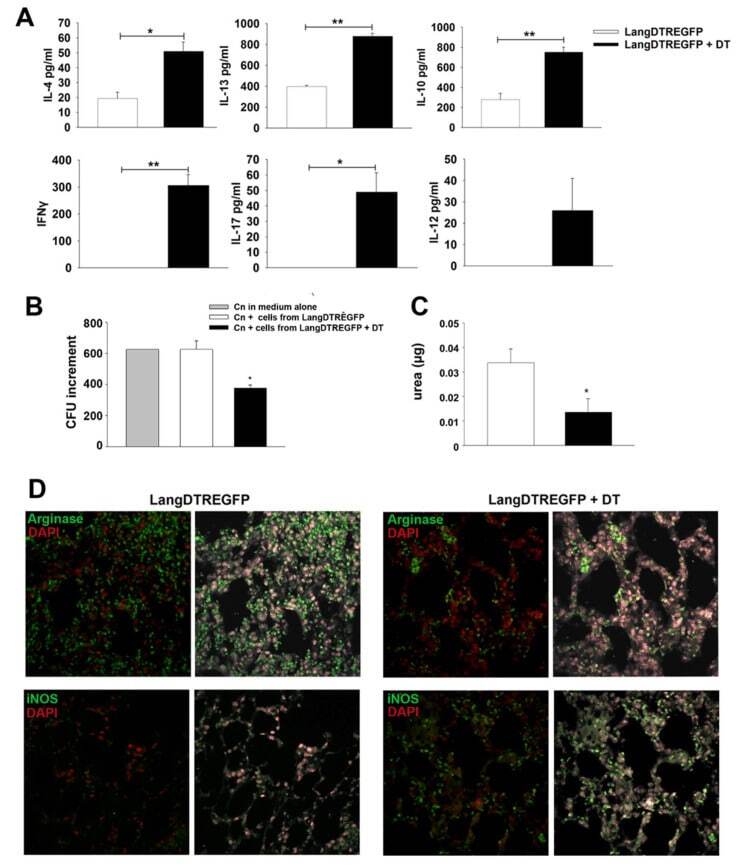
- Ex vivo cytokine production, fungal growth inhibition and arginase-1 or iNOS expression by lung cells from C. neoformans -infected mice. ( A ) Cytokine production by lung cell suspensions obtained from DT-treated (black bars) or untreated (white bars) LangDTREGFP mice at 7 days post infection. Bars represent cytokine levels (ELISA) in 24 h culture supernatants. * p < 0.05; ** p < 0.005. ( B ) In vitro C. neoformans growth assay using a yeast suspension (1 x 10 3 ) cultured in medium alone (gray bar) or in the presence of adherent lung cells from untreated (white bar) or DT-treated (black bar) 7-day-infected LangDTREGFP mice. Bars represent ratio of final vs initial CFU numbers in each culture condition. ( C ) Arginase activity from lysates of adherent cells cultured in ( B ), measured as urea production (ug of urea per ug of protein). * p < 0.05. Data are expressed as mean +- SEM. The data shown are pooled from two independent experiments ( n = 6 animals per group; samples from each animal were analyzed in triplicate ( A , C ) or duplicate ( B )). All data were analyzed with Student's t -test or ANOVA. ( D ) Confocal microscopy of representative lung sections from 7-day-infected mice ( n = 6) after immunostaining with fluorochrome-labeled specific antibodies to arginase-1 (upper panels) or iNOS (lower panels). 600x magnification. DT: diphtheria toxin; dpi: days post infection; CFU: colony-forming units.
- Conjugate
- Green dye