Explore
 Validate
 Learn
Learn Western blot
 Immunohistochemistry
Antibody data
- Antibody Data
- Antigen structure
- References [2]
- Comments [0]
- Validations
- Immunohistochemistry [1]
Submit
Validation data
Reference
Comment
Report error
- Product number
- M01106-2 - Provider product page

- Provider
- Boster Biological Technology
- Product name
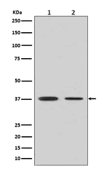
- Anti-Liver Arginase ARG1 Rabbit Monoclonal Antibody
- Antibody type
- Monoclonal
- Description
- Monoclonal antibody for Arginase 1/ARG1 detection. Host: Rabbit.Size: 100ug/vial. Tested applications: IP, IF, IHC, ICC, WB. Reactive species: Human Arginase 1/ARG1 information: Molecular Weight: 34735 MW; Subcellular Localization: Cytoplasm .
- Reactivity
- Human
- Host
- Rabbit
- Antibody clone number
- ABO-1
- Vial size
- 100ug/vial
- Concentration
- 0.5-1mg/ml, actual concentration vary by lot. Use suggested dilution ratio to decide dilution procedure.
- Storage
- At -20°C for one year. Avoid repeated freezing and thawing.
Submitted references A one-two punch strategy for diabetic wound management based on an antibiotic-hybrid biomineralized iron sulfide nanoparticle.
MiR196a-5p in extracellular vesicles released from human nasopharyngeal carcinoma enhance the phagocytosis and secretion of microglia by targeting ROCK1.
Deng S, Ou K, Zhang C, Yuan D, Cai X, Li F, Wang X, Yin J, Xu C, Li Y, Gong T
Acta biomaterialia 2024 Jun;181:333-346
Acta biomaterialia 2024 Jun;181:333-346
MiR196a-5p in extracellular vesicles released from human nasopharyngeal carcinoma enhance the phagocytosis and secretion of microglia by targeting ROCK1.
Chen P, Liu R, Yu Z, Cui G, Zong W, Wang M, Xie M, Qu W, Wang W, Luo X
Experimental cell research 2022 Feb 15;411(2):112988
Experimental cell research 2022 Feb 15;411(2):112988
No comments: Submit comment
Supportive validation
- Submitted by
- Boster Biological Technology (provider)
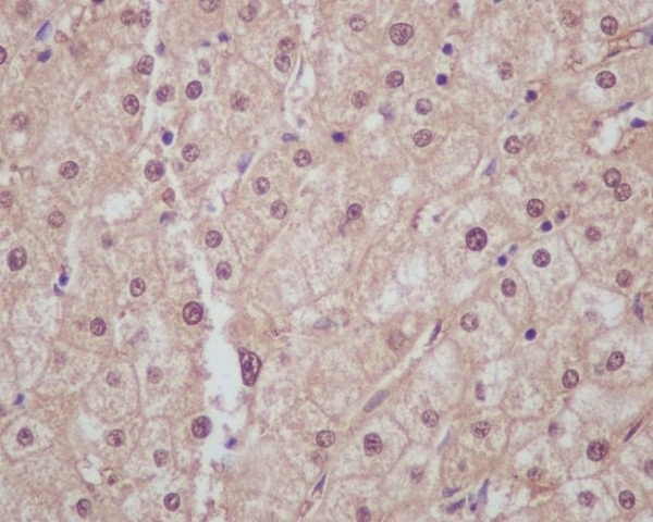
- Main image
- Experimental details
- Immunohistochemical analysis of paraffin-embedded human liver, using Liver Arginase Antibody(M01106-2)ARG1 was detected in paraffin-embedded tissue section. Heat mediated antigen retrieval was performed in citrate buffer (pH6, epitope retrieval solution) for 20 mins. The tissue section was blocked with 10% goat serum. The tissue section was then incubated with 1ug/ml rabbit anti-ARG1 Antibody (M01106-2)overnight at 4?? Biotinylated goat anti-rabbit IgG was used as secondary antibody and incubated for 30 minutes at 37?? The tissue section was developed using Strepavidin-Biotin-Complex (SABC)(Catalog # SA1022) with DAB as the chromogen.
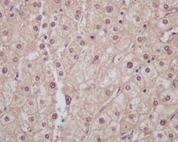
- Additional image