Explore
 Validate
 Learn
Learn Western blot
 Immunocytochemistry
Antibody data
- Antibody Data
- Antigen structure
- References [7]
- Comments [0]
- Validations
- Western blot [5]
- Immunoprecipitation [1]
- Immunohistochemistry [3]
- Flow cytometry [2]
Submit
Validation data
Reference
Comment
Report error
- Product number
- NBP1-32731 - Provider product page

- Provider
- Novus Biologicals
- Proper citation
- Novus Cat#NBP1-32731, RRID:AB_10003985
- Product name
- Rabbit Polyclonal Arginase 1/ARG1/liver Arginase Antibody
- Antibody type
- Polyclonal
- Description
- Immunogen affinity purified.
- Reactivity
- Human, Mouse, Rat
- Host
- Rabbit
- Isotype
- IgG
- Vial size
- 0.1 mg
- Concentration
- 1.0 mg/ml
- Storage
- Aliquot and store at -20C or -80C. Avoid freeze-thaw cycles.
Submitted references Effect of sex differences in treatment response to angioplasty in a murine arteriovenous fistula model.
Protective role of berberine in isoprenaline-induced cardiac fibrosis in rats.
A novel Microproteomic Approach Using Laser Capture Microdissection to Study Cellular Protrusions.
Regulatory role of IKKɑ in myocardial ischemia/reperfusion injury by the determination of M1 versus M2 polarization of macrophages.
Tumour-vasculature development via endothelial-to-mesenchymal transition after radiotherapy controls CD44v6(+) cancer cell and macrophage polarization.
Autophagy Sustains Pancreatic Cancer Growth through Both Cell-Autonomous and Nonautonomous Mechanisms.
Diabetes impairs wound healing by Dnmt1-dependent dysregulation of hematopoietic stem cells differentiation towards macrophages.
Cai C, Zhao C, Kilari S, Sharma A, Singh AK, Simeon ML, Misra A, Li Y, Misra S
American journal of physiology. Renal physiology 2020 Mar 1;318(3):F565-F575
American journal of physiology. Renal physiology 2020 Mar 1;318(3):F565-F575
Protective role of berberine in isoprenaline-induced cardiac fibrosis in rats.
Che Y, Shen DF, Wang ZP, Jin YG, Wu QQ, Wang SS, Yuan Y
BMC cardiovascular disorders 2019 Oct 15;19(1):219
BMC cardiovascular disorders 2019 Oct 15;19(1):219
A novel Microproteomic Approach Using Laser Capture Microdissection to Study Cellular Protrusions.
Gousset K, Gordon A, Kumar Kannan S, Tovar J
International journal of molecular sciences 2019 Mar 7;20(5)
International journal of molecular sciences 2019 Mar 7;20(5)
Regulatory role of IKKɑ in myocardial ischemia/reperfusion injury by the determination of M1 versus M2 polarization of macrophages.
Cao Y, Xu Y, Auchoybur ML, Chen W, He S, Qin W, Su C, Huang F, Qiu Z, Li L, Chen X
Journal of molecular and cellular cardiology 2018 Oct;123:1-12
Journal of molecular and cellular cardiology 2018 Oct;123:1-12
Tumour-vasculature development via endothelial-to-mesenchymal transition after radiotherapy controls CD44v6(+) cancer cell and macrophage polarization.
Choi SH, Kim AR, Nam JK, Kim JM, Kim JY, Seo HR, Lee HJ, Cho J, Lee YJ
Nature communications 2018 Nov 30;9(1):5108
Nature communications 2018 Nov 30;9(1):5108
Autophagy Sustains Pancreatic Cancer Growth through Both Cell-Autonomous and Nonautonomous Mechanisms.
Yang A, Herter-Sprie G, Zhang H, Lin EY, Biancur D, Wang X, Deng J, Hai J, Yang S, Wong KK, Kimmelman AC
Cancer discovery 2018 Mar;8(3):276-287
Cancer discovery 2018 Mar;8(3):276-287
Diabetes impairs wound healing by Dnmt1-dependent dysregulation of hematopoietic stem cells differentiation towards macrophages.
Yan J, Tie G, Wang S, Tutto A, DeMarco N, Khair L, Fazzio TG, Messina LM
Nature communications 2018 Jan 2;9(1):33
Nature communications 2018 Jan 2;9(1):33
No comments: Submit comment
Supportive validation
- Submitted by
- Novus Biologicals (provider)
- Main image
- Experimental details
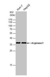
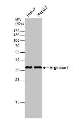
- Western Blot: Arginase 1/ARG1/liver Arginase Antibody [NBP1-32731] - Arginase 1 antibody detects Arginase 1 protein. Various whole cell extracts (30ug) were separated by 12% SDS-PAGE, and the membrane was blotted with Arginase 1 antibody diluted at 1:1000.
- Submitted by
- Novus Biologicals (provider)
- Main image
- Experimental details
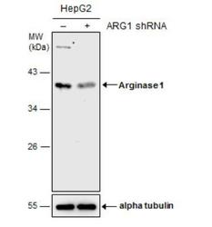
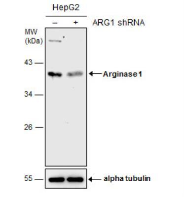
- Western Blot: Arginase 1/ARG1/liver Arginase Antibody [NBP1-32731] - Non-transfected (-) and transfected (+) HepG2 whole cell extracts (30 ug) were separated by 10% SDS-PAGE, and the membrane was blotted with Arginase 1 antibody.
- Submitted by
- Novus Biologicals (provider)
- Main image
- Experimental details
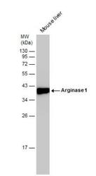
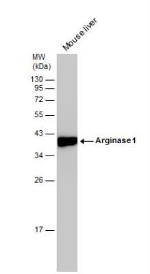
- Western Blot: Arginase 1/ARG1/liver Arginase Antibody [NBP1-32731] - Mouse tissue extract (50 ug) was separated by 12% SDS-PAGE, and the membrane was blotted with Arginase 1 antibody diluted at 1:10000.
- Submitted by
- Novus Biologicals (provider)
- Main image
- Experimental details
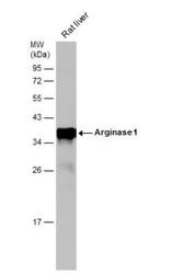
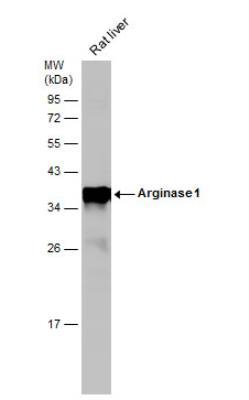
- Western Blot: Arginase 1/ARG1/liver Arginase Antibody [NBP1-32731] - Rat tissue extract (50 ug) was separated by 12% SDS-PAGE, and the membrane was blotted with Arginase 1 antibody diluted at 1:10000.
- Submitted by
- Novus Biologicals (provider)
- Main image
- Experimental details
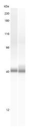
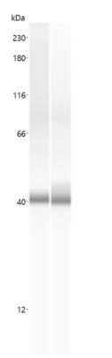
- Simple Western: Arginase 1/ARG1/liver Arginase Antibody [NBP1-32731] - Simple Western lane view shows a specific band for ARG1 in human and mouse Liver lysate using ARG1 antibody (NBP1-32731) at 25 ug/ml. This experiment was performed under reducing conditions using the 12-230 kDa separation system.
Supportive validation
- Submitted by
- Novus Biologicals (provider)
- Main image
- Experimental details
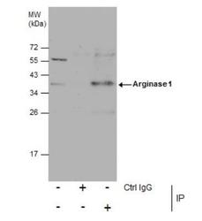
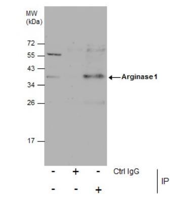
- Immunoprecipitation: Arginase 1/ARG1/liver Arginase Antibody [NBP1-32731] - Analysis was performed using Arginase 1 antibody EasyBlot anti-Rabbit IgG was used as a secondary reagent. Arginase 1 protein from HepG2 whole cell extracts using 5 ug of Arginase 1 antibody.
Supportive validation
- Submitted by
- Novus Biologicals (provider)
- Main image
- Experimental details
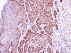
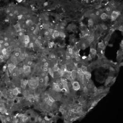
- Immunohistochemistry-Paraffin: Arginase 1/ARG1/liver Arginase Antibody [NBP1-32731] - Cal27 xenograft, using arginase I antibody at 1:500 dilution.
- Submitted by
- Novus Biologicals (provider)
- Main image
- Experimental details
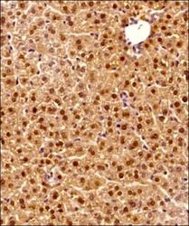
- Immunohistochemistry-Paraffin: Arginase 1/ARG1/liver Arginase Antibody [NBP1-32731] - Tissue section of the mouse liver using 1:200 dilution of ARG1 antibody (NBP1-32731). The signal was developed using HRP-DAB method which followed counterstaining of the cells with hematoxylin.

- Submitted by
- Novus Biologicals (provider)
- Main image
- Experimental details
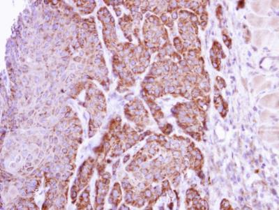
- Immunohistochemistry-Paraffin: Arginase 1/ARG1/liver Arginase Antibody [NBP1-32731] - Hepatocellular carcinoma stained with Arginase1 1:100, pH9 antigen retrieval. Image from verified customer review.

Supportive validation
- Submitted by
- Novus Biologicals (provider)
- Main image
- Experimental details
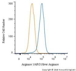
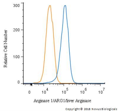
- Flow Cytometry: Arginase 1/ARG1/liver Arginase Antibody [NBP1-32731] - An intracellular stain was performed on HepG2 with NBP1-32731 and a matched isotype control. Cells were fixed with 4% PFA and then permeablized with 0.1% saponin. Cells were incubated in an antibody dilution of 5 ug/mL for 30 minutes at room temperature, followed by Rabbit IgG (H+L) Cross-Adsorbed Secondary Antibody.
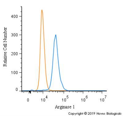
- Submitted by
- Novus Biologicals (provider)
- Main image
- Experimental details
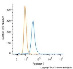
- Flow Cytometry: Arginase 1/ARG1/liver Arginase Antibody [NBP1-32731] - An intracellular stain was performed on RH30 cells with Arginase 1 Antibody NBP1-32731 (blue) and a matched isotype control (orange). Cells were fixed with 4% PFA and then permeabilized with 0.1% saponin. Cells were incubated in an antibody dilution of 5.0 ug/mL for 30 minutes at room temperature, followed by Rabbit IgG (H+L) Cross-Adsorbed Secondary Antibody.