 Explore
 Validate
 Learn
Learn Western blot
Antibody data
- Antibody Data
- Antigen structure
- References [3]
- Comments [0]
- Validations
- Western blot [2]
- Immunohistochemistry [3]
Submit
Validation data
Reference
Comment
Report error
- Product number
- NBP1-87455 - Provider product page

- Provider
- Novus Biologicals
- Proper citation
- Novus Cat#NBP1-87455, RRID:AB_11040973
- Product name
- Rabbit Polyclonal Arginase 1/ARG1/liver Arginase Antibody
- Antibody type
- Polyclonal
- Description
- Immunogen affinity purified. Specificity of human Arginase 1/ARG1/liver Arginase antibody verified on a Protein Array containing target protein plus 383 other non-specific proteins.
- Reactivity
- Human, Mouse
- Host
- Rabbit
- Isotype
- IgG
- Vial size
- 0.1 ml
- Storage
- Store at 4C short term. Aliquot and store at -20C long term. Avoid freeze-thaw cycles.
Submitted references Interleukin-6/interleukin-21 signaling axis is critical in the pathogenesis of pulmonary arterial hypertension.
Initial quantitative proteomic map of 28 mouse tissues using the SILAC mouse.
Expression patterns of the immunomodulatory enzyme arginase 1 in blood, lymph nodes and tumor tissue of early-stage breast cancer patients.
Hashimoto-Kataoka T, Hosen N, Sonobe T, Arita Y, Yasui T, Masaki T, Minami M, Inagaki T, Miyagawa S, Sawa Y, Murakami M, Kumanogoh A, Yamauchi-Takihara K, Okumura M, Kishimoto T, Komuro I, Shirai M, Sakata Y, Nakaoka Y
Proceedings of the National Academy of Sciences of the United States of America 2015 May 19;112(20):E2677-86
Proceedings of the National Academy of Sciences of the United States of America 2015 May 19;112(20):E2677-86
Initial quantitative proteomic map of 28 mouse tissues using the SILAC mouse.
Geiger T, Velic A, Macek B, Lundberg E, Kampf C, Nagaraj N, Uhlen M, Cox J, Mann M
Molecular & cellular proteomics : MCP 2013 Jun;12(6):1709-22
Molecular & cellular proteomics : MCP 2013 Jun;12(6):1709-22
Expression patterns of the immunomodulatory enzyme arginase 1 in blood, lymph nodes and tumor tissue of early-stage breast cancer patients.
de Boniface J, Mao Y, Schmidt-Mende J, Kiessling R, Poschke I
Oncoimmunology 2012 Nov 1;1(8):1305-1312
Oncoimmunology 2012 Nov 1;1(8):1305-1312
No comments: Submit comment
Supportive validation
- Submitted by
- Novus Biologicals (provider)
- Main image

- Experimental details
- Western Blot: Arginase 1/ARG1/liver Arginase Antibody [NBP1-87455] - Lane 1: Marker [kDa] 230, 130, 95, 72, 56, 36, 28, 17, 11. Lane 2: Human cell line RT-4. Lane 3: Human cell line U-251MG sp. Lane 4: Human plasma (IgG/HSA depleted). Lane 5: Human liver tissue

- Submitted by
- Novus Biologicals (provider)
- Main image

- Experimental details
- Western Blot: Arginase 1/ARG1/liver Arginase Antibody [NBP1-87455] - Analysis using Anti-ARG1 antibody NBP1-87455 (A) shows similar pattern to independent antibody NBP1-87490 (B).

Supportive validation
- Submitted by
- Novus Biologicals (provider)
- Main image
- Experimental details
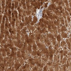
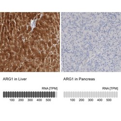
- Immunohistochemistry-Paraffin: Arginase 1/ARG1/liver Arginase Antibody [NBP1-87455] - Staining of human liver shows high expression.

- Submitted by
- Novus Biologicals (provider)
- Main image
- Experimental details
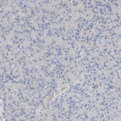
- Immunohistochemistry-Paraffin: Arginase 1/ARG1/liver Arginase Antibody [NBP1-87455] - Staining of human pancreas shows low expression as expected.

- Submitted by
- Novus Biologicals (provider)
- Main image
- Experimental details
- Immunohistochemistry-Paraffin: Arginase 1/ARG1/liver Arginase Antibody [NBP1-87455] - Staining in human liver and pancreas tissues using anti-ARG1 antibody. Corresponding ARG1 RNA-seq data are presented for the same tissues.
