Explore
 Validate
 Learn
Learn Western blot
 Immunohistochemistry
Antibody data
- Antibody Data
- Antigen structure
- References [17]
- Comments [0]
- Validations
- Immunohistochemistry [1]
Submit
Validation data
Reference
Comment
Report error
- Product number
- HPA003595 - Provider product page

- Provider
- Atlas Antibodies
- Proper citation
- Atlas Antibodies Cat#HPA003595, RRID:AB_1078190
- Product name
- Anti-ARG1
- Antibody type
- Polyclonal
- Description
- Polyclonal Antibody against Human ARG1, Gene description: arginase 1, Validated applications: IHC, WB, Uniprot ID: P05089, Storage: Store at +4°C for short term storage. Long time storage is recommended at -20°C.
- Reactivity
- Human
- Host
- Rabbit
- Conjugate
- Unconjugated
- Isotype
- IgG
- Vial size
- 100 µl
- Concentration
- 0.2 mg/ml
- Storage
- Store at +4°C for short term storage. Long time storage is recommended at -20°C.
- Handling
- The antibody solution should be gently mixed before use.
Submitted references Liraglutide Therapy in Obese Patients Alters Macrophage Phenotype and Decreases Their Tumor Necrosis Factor Alpha Release and Oxidative Stress Markers—A Pilot Study
Characterisation of Expression the Arginine Pathway Enzymes in Childhood Brain Tumours to Determine Susceptibility to Therapeutic Arginine Depletion
A Novel NIPBL-NACC1 Gene Fusion Is Characteristic of the Cholangioblastic Variant of Intrahepatic Cholangiocarcinoma
Evaluation of Glycosylated PTGS2 in Colorectal Cancer for NSAIDS-Based Adjuvant Therapy
LmjF.22.0810 from Leishmania major Modulates the Th2-Type Immune Response and Is Involved in Leishmaniasis Outcome
Transient Support from Fibroblasts is Sufficient to Drive Functional Vascularization in Engineered Tissues
M2-Polarized Macrophages Determine Human Cutaneous Lesions in Lacaziosis
High-density neutrophils in MGUS and multiple myeloma are dysfunctional and immune-suppressive due to increased STAT3 downstream signaling
Alpha-Fetoprotein-Producing Early Gastric Cancer with Intramucosal Hepatoid and Fetal Enteric Differentiation.
Plasticity of High-Density Neutrophils in Multiple Myeloma is Associated with Increased Autophagy Via STAT3
The prognostic value of the myeloid-mediated immunosuppression marker Arginase-1 in classic Hodgkin lymphoma
Arginase regulates red blood cell nitric oxide synthase and export of cardioprotective nitric oxide bioactivity
Local Arginase Inhibition during Early Reperfusion Mediates Cardioprotection via Increased Nitric Oxide Production
Arginase Inhibition Improves Endothelial Function in Patients With Coronary Artery Disease and Type 2 Diabetes Mellitus
Arginase inhibition restores in vivo coronary microvascular function in type 2 diabetic rats
Arginase-1
A Quantitative Proteomic Approach for Identification of Potential Biomarkers in Hepatocellular Carcinoma
Bułdak Ł, Bołdys A, Skudrzyk E, Machnik G, Okopień B
Metabolites 2024;14(10):554
Metabolites 2024;14(10):554
Characterisation of Expression the Arginine Pathway Enzymes in Childhood Brain Tumours to Determine Susceptibility to Therapeutic Arginine Depletion
Bishop E, Dimitrova M, Froggatt A, Estevez-Cebrero M, Storer L, Mussai F, Paine S, Grundy R, Dandapani M, Prodam F
BioMed Research International 2022;2022(1)
BioMed Research International 2022;2022(1)
A Novel NIPBL-NACC1 Gene Fusion Is Characteristic of the Cholangioblastic Variant of Intrahepatic Cholangiocarcinoma
Argani P, Palsgrove D, Anders R, Smith S, Saoud C, Kwon R, Voltaggio L, Assarzadegan N, Oshima K, Rooper L, Matoso A, Zhang L, Cantarel B, Gagan J, Antonescu C
American Journal of Surgical Pathology 2021;45(11):1550-1560
American Journal of Surgical Pathology 2021;45(11):1550-1560
Evaluation of Glycosylated PTGS2 in Colorectal Cancer for NSAIDS-Based Adjuvant Therapy
Venè R, Costa D, Augugliaro R, Carlone S, Scabini S, Casoni Pattacini G, Boggio M, Zupo S, Grillo F, Mastracci L, Pitto F, Minghelli S, Ferrari N, Tosetti F, Romairone E, Mingari M, Poggi A, Benelli R
Cells 2020;9(3):683
Cells 2020;9(3):683
LmjF.22.0810 from Leishmania major Modulates the Th2-Type Immune Response and Is Involved in Leishmaniasis Outcome
Vacas A, Fernández-Rubio C, Larrea E, Peña-Guerrero J, Nguewa P
Biomedicines 2020;8(11):452
Biomedicines 2020;8(11):452
Transient Support from Fibroblasts is Sufficient to Drive Functional Vascularization in Engineered Tissues
Song H, Lammers A, Sundaram S, Rubio L, Chen A, Li L, Eyckmans J, Bhatia S, Chen C
Advanced Functional Materials 2020;30(48)
Advanced Functional Materials 2020;30(48)
M2-Polarized Macrophages Determine Human Cutaneous Lesions in Lacaziosis
Barboza T, Sotto M, Kanashiro-Galo L, de Brito A, Duarte M, Quaresma J, Pagliari C
Mycopathologia 2020;185(3):477-483
Mycopathologia 2020;185(3):477-483
High-density neutrophils in MGUS and multiple myeloma are dysfunctional and immune-suppressive due to increased STAT3 downstream signaling
Romano A, Parrinello N, Simeon V, Puglisi F, La Cava P, Bellofiore C, Giallongo C, Camiolo G, D’Auria F, Grieco V, Larocca F, Barbato A, Cambria D, La Spina E, Tibullo D, Palumbo G, Conticello C, Musto P, Di Raimondo F
Scientific Reports 2020;10(1)
Scientific Reports 2020;10(1)
Alpha-Fetoprotein-Producing Early Gastric Cancer with Intramucosal Hepatoid and Fetal Enteric Differentiation.
Iwaya M, Riddell R, Asano K, Kobayashi K, Uehara T, Ota H
Case reports in gastroenterology 2020 May-Aug;14(2):426-435
Case reports in gastroenterology 2020 May-Aug;14(2):426-435
Plasticity of High-Density Neutrophils in Multiple Myeloma is Associated with Increased Autophagy Via STAT3
Puglisi F, Parrinello N, Giallongo C, Cambria D, Camiolo G, Bellofiore C, Conticello C, Del Fabro V, Leotta V, Markovic U, Sapienza G, Barbato A, Scalese S, Tibullo D, Brundo M, Palumbo G, Di Raimondo F, Romano A
International Journal of Molecular Sciences 2019;20(14):3548
International Journal of Molecular Sciences 2019;20(14):3548
The prognostic value of the myeloid-mediated immunosuppression marker Arginase-1 in classic Hodgkin lymphoma
Romano A, Parrinello N, Vetro C, Tibullo D, Giallongo C, La Cava P, Chiarenza A, Motta G, Caruso A, Villari L, Tripodo C, Cosentino S, Ippolito M, Consoli U, Gallamini A, Pileri S, Di Raimondo F
Oncotarget 2016;7(41):67333-67346
Oncotarget 2016;7(41):67333-67346
Arginase regulates red blood cell nitric oxide synthase and export of cardioprotective nitric oxide bioactivity
Yang J, Gonon A, Sjöquist P, Lundberg J, Pernow J
Proceedings of the National Academy of Sciences 2013;110(37):15049-15054
Proceedings of the National Academy of Sciences 2013;110(37):15049-15054
Local Arginase Inhibition during Early Reperfusion Mediates Cardioprotection via Increased Nitric Oxide Production
Gallyas F, Gonon A, Jung C, Katz A, Westerblad H, Shemyakin A, Sjöquist P, Lundberg J, Pernow J
PLoS ONE 2012;7(7):e42038
PLoS ONE 2012;7(7):e42038
Arginase Inhibition Improves Endothelial Function in Patients With Coronary Artery Disease and Type 2 Diabetes Mellitus
Shemyakin A, Kövamees O, Rafnsson A, Böhm F, Svenarud P, Settergren M, Jung C, Pernow J
Circulation 2012;126(25):2943-2950
Circulation 2012;126(25):2943-2950
Arginase inhibition restores in vivo coronary microvascular function in type 2 diabetic rats
Grönros J, Jung C, Lundberg J, Cerrato R, Östenson C, Pernow J
American Journal of Physiology-Heart and Circulatory Physiology 2011;300(4):H1174-H1181
American Journal of Physiology-Heart and Circulatory Physiology 2011;300(4):H1174-H1181
Arginase-1
Yan B, Gong C, Song J, Krausz T, Tretiakova M, Hyjek E, Al-Ahmadie H, Alves V, Xiao S, Anders R, Hart J
American Journal of Surgical Pathology 2010;34(8):1147-1154
American Journal of Surgical Pathology 2010;34(8):1147-1154
A Quantitative Proteomic Approach for Identification of Potential Biomarkers in Hepatocellular Carcinoma
Chaerkady R, Harsha H, Nalli A, Gucek M, Vivekanandan P, Akhtar J, Cole R, Simmers J, Schulick R, Singh S, Torbenson M, Pandey A, Thuluvath P
Journal of Proteome Research 2008;7(10):4289-4298
Journal of Proteome Research 2008;7(10):4289-4298
No comments: Submit comment
Supportive validation
- Submitted by
- Atlas Antibodies (provider)
- Enhanced method
- Orthogonal validation
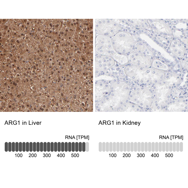
- Main image

- Experimental details
- Immunohistochemistry analysis in human liver and kidney tissues using HPA003595 antibody. Corresponding ARG1 RNA-seq data are presented for the same tissues.
- Sample type
- Human
- Protocol
- Protocol