Explore
 Validate
 Learn
Learn Western blot
 Immunoprecipitation
Antibody data
- Antibody Data
- Antigen structure
- References [7]
- Comments [0]
- Validations
- Western blot [2]
Submit
Validation data
Reference
Comment
Report error
- Product number
- AF5868 - Provider product page

- Provider
- R&D Systems
- Product name
- Human/Mouse/Rat Arginase 1/ARG1 Antibody
- Antibody type
- Polyclonal
- Description
- Antigen Affinity-purified. Detects human, mouse, and rat Arginase 1/ARG1 in direct ELISAs and Western blots.
- Reactivity
- Human, Mouse, Rat
- Host
- Sheep
- Conjugate
- Unconjugated
- Antigen sequence
P05089- Isotype
- IgG
- Vial size
- 100 ug
- Concentration
- LYOPH
- Storage
- Use a manual defrost freezer and avoid repeated freeze-thaw cycles. 12 months from date of receipt, -20 to -70 °C as supplied. 1 month, 2 to 8 °C under sterile conditions after reconstitution. 6 months, -20 to -70 °C under sterile conditions after reconstitution.
Submitted references The APMAP interactome reveals new modulators of APP processing and beta-amyloid production that are altered in Alzheimer's disease.
Extracellular vesicles secreted by hypoxia pre-challenged mesenchymal stem cells promote non-small cell lung cancer cell growth and mobility as well as macrophage M2 polarization via miR-21-5p delivery.
Arginase1 Deficiency in Monocytes/Macrophages Upregulates Inducible Nitric Oxide Synthase To Promote Cutaneous Contact Hypersensitivity.
Assessment of Preclinical Liver and Skeletal Muscle Biomarkers Following Clofibrate Administration in Wistar Rats.
Candida albicans Chitin Increases Arginase-1 Activity in Human Macrophages, with an Impact on Macrophage Antimicrobial Functions.
IL-33 contributes to sepsis-induced long-term immunosuppression by expanding the regulatory T cell population.
DAMP signaling is a key pathway inducing immune modulation after brain injury.
Gerber H, Mosser S, Boury-Jamot B, Stumpe M, Piersigilli A, Goepfert C, Dengjel J, Albrecht U, Magara F, Fraering PC
Acta neuropathologica communications 2019 Jan 31;7(1):13
Acta neuropathologica communications 2019 Jan 31;7(1):13
Extracellular vesicles secreted by hypoxia pre-challenged mesenchymal stem cells promote non-small cell lung cancer cell growth and mobility as well as macrophage M2 polarization via miR-21-5p delivery.
Ren W, Hou J, Yang C, Wang H, Wu S, Wu Y, Zhao X, Lu C
Journal of experimental & clinical cancer research : CR 2019 Feb 8;38(1):62
Journal of experimental & clinical cancer research : CR 2019 Feb 8;38(1):62
Arginase1 Deficiency in Monocytes/Macrophages Upregulates Inducible Nitric Oxide Synthase To Promote Cutaneous Contact Hypersensitivity.
Suwanpradid J, Shih M, Pontius L, Yang B, Birukova A, Guttman-Yassky E, Corcoran DL, Que LG, Tighe RM, MacLeod AS
Journal of immunology (Baltimore, Md. : 1950) 2017 Sep 1;199(5):1827-1834
Journal of immunology (Baltimore, Md. : 1950) 2017 Sep 1;199(5):1827-1834
Assessment of Preclinical Liver and Skeletal Muscle Biomarkers Following Clofibrate Administration in Wistar Rats.
Maliver P, Festag M, Bennecke M, Christen F, Bánfai B, Lenz B, Winter M
Toxicologic pathology 2017 Jun;45(4):506-525
Toxicologic pathology 2017 Jun;45(4):506-525
Candida albicans Chitin Increases Arginase-1 Activity in Human Macrophages, with an Impact on Macrophage Antimicrobial Functions.
Wagener J, MacCallum DM, Brown GD, Gow NA
mBio 2017 Jan 24;8(1)
mBio 2017 Jan 24;8(1)
IL-33 contributes to sepsis-induced long-term immunosuppression by expanding the regulatory T cell population.
Nascimento DC, Melo PH, Piñeros AR, Ferreira RG, Colón DF, Donate PB, Castanheira FV, Gozzi A, Czaikoski PG, Niedbala W, Borges MC, Zamboni DS, Liew FY, Cunha FQ, Alves-Filho JC
Nature communications 2017 Apr 4;8:14919
Nature communications 2017 Apr 4;8:14919
DAMP signaling is a key pathway inducing immune modulation after brain injury.
Liesz A, Dalpke A, Mracsko E, Antoine DJ, Roth S, Zhou W, Yang H, Na SY, Akhisaroglu M, Fleming T, Eigenbrod T, Nawroth PP, Tracey KJ, Veltkamp R
The Journal of neuroscience : the official journal of the Society for Neuroscience 2015 Jan 14;35(2):583-98
The Journal of neuroscience : the official journal of the Society for Neuroscience 2015 Jan 14;35(2):583-98
No comments: Submit comment
Supportive validation
- Submitted by
- R&D Systems (provider)
- Main image
- Experimental details
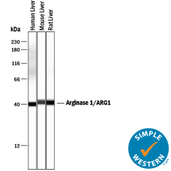
- Detection of Human, Mouse, and Rat Arginase 1/ARG1 by Simple WesternTM. Simple Western lane view shows lysates of human liver tissue, mouse liver tissue, and rat liver tissue, loaded at 0.2 mg/mL. A specific band was detected for Arginase 1/ARG1 at approximately 41 kDa (as indicated) using 2.5 µg/mL of Sheep Anti-Human/Mouse/Rat Arginase 1/ARG1 Antigen Affinity-purified Polyclonal Antibody (Catalog # AF5868) followed by 1:50 dilution of HRP-conjugated Anti-Sheep IgG Secondary Antibody (Catalog # HAF016). This experiment was conducted under reducing conditions and using the 12-230 kDa separation system.

- Submitted by
- R&D Systems (provider)
- Main image

- Experimental details
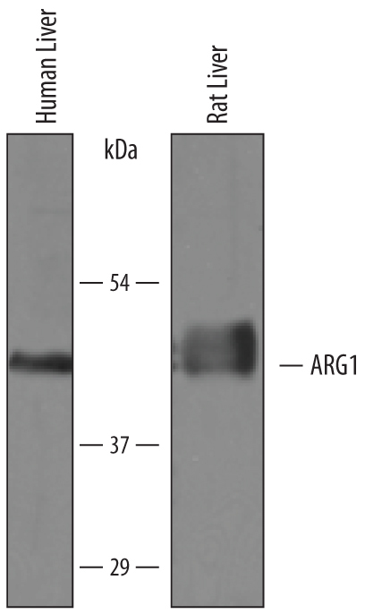
- Detection of Human and Rat Arginase 1/ARG1 by Western Blot. Western blot shows lysates of human liver tissue and rat liver tissue. PVDF membrane was probed with 0.25 µg/mL of Sheep Anti-Human/Mouse/Rat Arginase 1/ARG1 Antigen Affinity-purified Polyclonal Antibody (Catalog # AF5868) followed by HRP-conjugated Anti-Sheep IgG Secondary Antibody (Catalog # HAF016). A specific band was detected for Arginase 1/ARG1 at approximately 41 kDa (as indicated). This experiment was conducted under reducing conditions and using Immunoblot Buffer Group 8.