Explore
 Validate
 Learn
Learn Flow cytometry
Antibody data
- Antibody Data
- Antigen structure
- References [12]
- Comments [0]
- Validations
- Flow cytometry [2]
Submit
Validation data
Reference
Comment
Report error
- Product number
- IC5868P - Provider product page

- Provider
- R&D Systems
- Product name
- Human/Mouse Arginase 1/ARG1 PE-conjugated Antibody
- Antibody type
- Polyclonal
- Description
- Antigen Affinity-purified. Detects human, mouse, and rat Arginase 1/ARG1 in direct ELISAs and Western blots.
- Reactivity
- Human, Mouse
- Host
- Sheep
- Antigen sequence
P05089- Isotype
- IgG
- Vial size
- 100 Tests
- Storage
- Protect from light. Do not freeze. 12 months from date of receipt, 2 to 8 °C as supplied.
Submitted references Concerted IL-25R and IL-4Rα signaling drive innate type 2 effector immunity for optimal helminth expulsion.
Sitravatinib potentiates immune checkpoint blockade in refractory cancer models.
Temporal changes in macrophage phenotype after peripheral nerve injury.
Chloroquine modulates antitumor immune response by resetting tumor-associated macrophages toward M1 phenotype.
Therapeutic benefits of phosphodiesterase 4B inhibition after traumatic brain injury.
IL-4 up-regulates cyclooxygenase-1 expression in macrophages.
Dysferlinopathy Promotes an Intramuscle Expansion of Macrophages with a Cyto-Destructive Phenotype.
Infection-adapted emergency hematopoiesis promotes visceral leishmaniasis.
Obesity triggers enhanced MDSC accumulation in murine renal tumors via elevated local production of CCL2.
Protective Role of STAT6 in Basophil-Dependent Prurigo-like Allergic Skin Inflammation.
IL-34 mediates acute kidney injury and worsens subsequent chronic kidney disease.
Anti-melanoma vaccines engineered to simultaneously modulate cytokine priming and silence PD-L1 characterized using ex vivo myeloid-derived suppressor cells as a readout of therapeutic efficacy.
Smith KA, Löser S, Varyani F, Harcus Y, McSorley HJ, McKenzie AN, Maizels RM
eLife 2018 Sep 21;7
eLife 2018 Sep 21;7
Sitravatinib potentiates immune checkpoint blockade in refractory cancer models.
Du W, Huang H, Sorrelle N, Brekken RA
JCI insight 2018 Nov 2;3(21)
JCI insight 2018 Nov 2;3(21)
Temporal changes in macrophage phenotype after peripheral nerve injury.
Tomlinson JE, Žygelytė E, Grenier JK, Edwards MG, Cheetham J
Journal of neuroinflammation 2018 Jun 15;15(1):185
Journal of neuroinflammation 2018 Jun 15;15(1):185
Chloroquine modulates antitumor immune response by resetting tumor-associated macrophages toward M1 phenotype.
Chen D, Xie J, Fiskesund R, Dong W, Liang X, Lv J, Jin X, Liu J, Mo S, Zhang T, Cheng F, Zhou Y, Zhang H, Tang K, Ma J, Liu Y, Huang B
Nature communications 2018 Feb 28;9(1):873
Nature communications 2018 Feb 28;9(1):873
Therapeutic benefits of phosphodiesterase 4B inhibition after traumatic brain injury.
Wilson NM, Gurney ME, Dietrich WD, Atkins CM
PloS one 2017;12(5):e0178013
PloS one 2017;12(5):e0178013
IL-4 up-regulates cyclooxygenase-1 expression in macrophages.
Shay AE, Diwakar BT, Guan BJ, Narayan V, Urban JF Jr, Prabhu KS
The Journal of biological chemistry 2017 Sep 1;292(35):14544-14555
The Journal of biological chemistry 2017 Sep 1;292(35):14544-14555
Dysferlinopathy Promotes an Intramuscle Expansion of Macrophages with a Cyto-Destructive Phenotype.
Baek JH, Many GM, Evesson FJ, Kelley VR
The American journal of pathology 2017 Jun;187(6):1245-1257
The American journal of pathology 2017 Jun;187(6):1245-1257
Infection-adapted emergency hematopoiesis promotes visceral leishmaniasis.
Abidin BM, Hammami A, Stäger S, Heinonen KM
PLoS pathogens 2017 Aug;13(8):e1006422
PLoS pathogens 2017 Aug;13(8):e1006422
Obesity triggers enhanced MDSC accumulation in murine renal tumors via elevated local production of CCL2.
Hale M, Itani F, Buchta CM, Wald G, Bing M, Norian LA
PloS one 2015;10(3):e0118784
PloS one 2015;10(3):e0118784
Protective Role of STAT6 in Basophil-Dependent Prurigo-like Allergic Skin Inflammation.
Hashimoto T, Satoh T, Yokozeki H
Journal of immunology (Baltimore, Md. : 1950) 2015 May 15;194(10):4631-40
Journal of immunology (Baltimore, Md. : 1950) 2015 May 15;194(10):4631-40
IL-34 mediates acute kidney injury and worsens subsequent chronic kidney disease.
Baek JH, Zeng R, Weinmann-Menke J, Valerius MT, Wada Y, Ajay AK, Colonna M, Kelley VR
The Journal of clinical investigation 2015 Aug 3;125(8):3198-214
The Journal of clinical investigation 2015 Aug 3;125(8):3198-214
Anti-melanoma vaccines engineered to simultaneously modulate cytokine priming and silence PD-L1 characterized using ex vivo myeloid-derived suppressor cells as a readout of therapeutic efficacy.
Liechtenstein T, Perez-Janices N, Blanco-Luquin I, Goyvaerts C, Schwarze J, Dufait I, Lanna A, Ridder M, Guerrero-Setas D, Breckpot K, Escors D
Oncoimmunology 2014;3(7):e945378
Oncoimmunology 2014;3(7):e945378
No comments: Submit comment
Supportive validation
- Submitted by
- R&D Systems (provider)
- Main image
- Experimental details
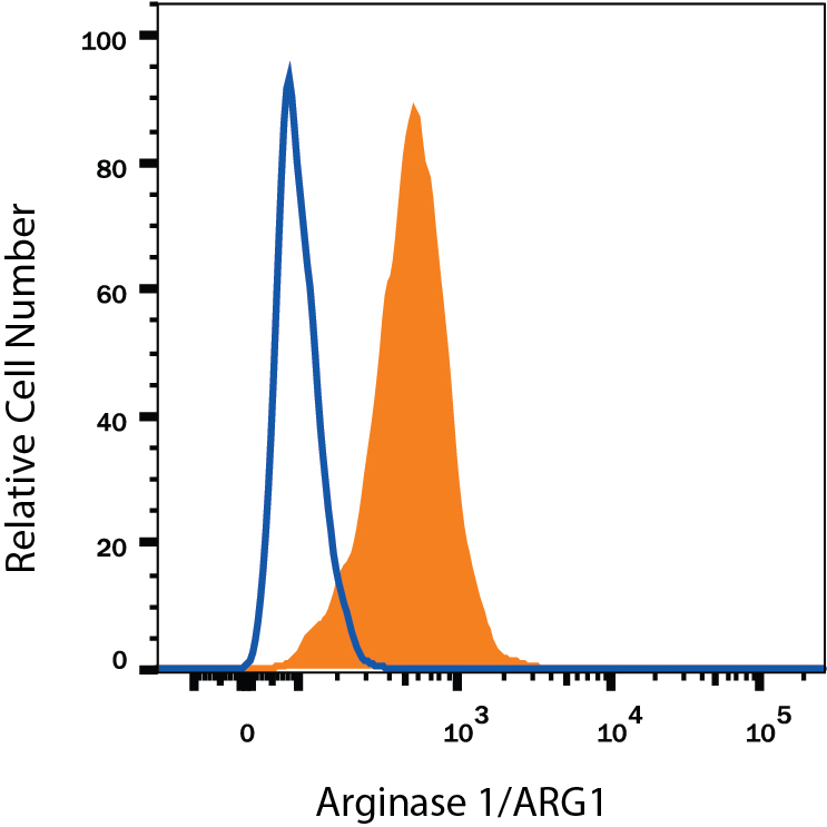
- Detection of Arginase 1/ARG1 in Hepa 1-6 Mouse Cell Line by Flow Cytometry. Hepa 1-6 mouse hepatoma cell line was stained with Sheep Anti-Human/Mouse Arginase 1/ARG1 PE-conjugated Antigen Affinity-purified Polyclonal Antibody (Catalog # IC5868P, filled histogram) or isotype control antibody (Catalog # IC016P, open histogram). To facilitate intracellular staining, cells were fixed with Flow Cytometry Fixation Buffer (Catalog # FC004) and permeabilized with Flow Cytometry Permeabilization/Wash Buffer I (Catalog # FC005). View our protocol for Staining Intracellular Molecules.
- Submitted by
- R&D Systems (provider)
- Main image

- Experimental details
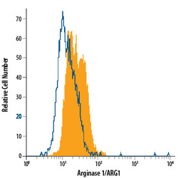
- Detection of Arginase 1/ARG1 in HepG2 Human Cell Line by Flow Cytometry. HepG2 human hepatocellular carcinoma cell line was stained with Sheep Anti-Human/Mouse Arginase 1/ARG1 PE-conjugated Antigen Affinity-purified Polyclonal Antibody (Catalog # IC5868P, filled histogram) or isotype control antibody (Catalog # IC016P, open histogram). To facilitate intracellular staining, cells were fixed with Flow Cytometry Fixation Buffer (Catalog # FC004) and permeabilized with Flow Cytometry Permeabilization/Wash Buffer I (Catalog # FC005). View our protocol for Staining Intracellular Molecules.