 Explore
 Validate
 Learn
Learn Western blot
Antibody data
- Antibody Data
- Antigen structure
- References [6]
- Comments [0]
- Validations
- Western blot [2]
- Immunocytochemistry [2]
- Immunohistochemistry [2]
- Other assay [3]
Submit
Validation data
Reference
Comment
Report error
- Product number
- 40-6100 - Provider product page

- Provider
- Invitrogen Antibodies
- Product name
- Occludin Polyclonal Antibody
- Antibody type
- Polyclonal
- Antigen
- Synthetic peptide
- Reactivity
- Human
- Host
- Rabbit
- Isotype
- IgG
- Vial size
- 100 µg
- Concentration
- 0.25 mg/mL
- Storage
- -20°C
Submitted references Occludin is a target of Src kinase and promotes lipid secretion by binding to BTN1a1 and XOR.
Effect of Bacillus subtilis Strains on Intestinal Barrier Function and Inflammatory Response.
Beta-catenin signaling regulates barrier-specific gene expression in circumventricular organ and ocular vasculatures.
Norrin/Frizzled4 signaling in retinal vascular development and blood brain barrier plasticity.
Anti-inflammatory and antioxidant effects of SERPINA3K in the retina.
Preconditioning with a TLR2 specific ligand increases resistance to cerebral ischemia/reperfusion injury.
Lu Y, Zhou T, Xu C, Wang R, Feng D, Li J, Wang X, Kong Y, Hu G, Kong X, Lu P
PLoS biology 2022 Jan;20(1):e3001518
PLoS biology 2022 Jan;20(1):e3001518
Effect of Bacillus subtilis Strains on Intestinal Barrier Function and Inflammatory Response.
Rhayat L, Maresca M, Nicoletti C, Perrier J, Brinch KS, Christian S, Devillard E, Eckhardt E
Frontiers in immunology 2019;10:564
Frontiers in immunology 2019;10:564
Beta-catenin signaling regulates barrier-specific gene expression in circumventricular organ and ocular vasculatures.
Wang Y, Sabbagh MF, Gu X, Rattner A, Williams J, Nathans J
eLife 2019 Apr 1;8
eLife 2019 Apr 1;8
Norrin/Frizzled4 signaling in retinal vascular development and blood brain barrier plasticity.
Wang Y, Rattner A, Zhou Y, Williams J, Smallwood PM, Nathans J
Cell 2012 Dec 7;151(6):1332-44
Cell 2012 Dec 7;151(6):1332-44
Anti-inflammatory and antioxidant effects of SERPINA3K in the retina.
Zhang B, Hu Y, Ma JX
Investigative ophthalmology & visual science 2009 Aug;50(8):3943-52
Investigative ophthalmology & visual science 2009 Aug;50(8):3943-52
Preconditioning with a TLR2 specific ligand increases resistance to cerebral ischemia/reperfusion injury.
Hua F, Ma J, Ha T, Kelley J, Williams DL, Kao RL, Kalbfleisch JH, Browder IW, Li C
Journal of neuroimmunology 2008 Aug 13;199(1-2):75-82
Journal of neuroimmunology 2008 Aug 13;199(1-2):75-82
No comments: Submit comment
Supportive validation
- Submitted by
- Invitrogen Antibodies (provider)
- Main image

- Experimental details
- Western blot analysis of Caco-2 cell lysates using Zymed Rb anti-Occludin (N-term) (Product # 40-6100).

- Submitted by
- Invitrogen Antibodies (provider)
- Main image

- Experimental details
- Western blot analysis of Caco-2 cell lysates using Zymed Rb anti-Occludin (N-term) (Product # 40-6100).

Supportive validation
- Submitted by
- Invitrogen Antibodies (provider)
- Main image

- Experimental details
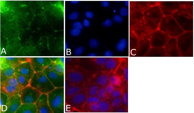
- Indirect immunofluorescent staining of Caco-2 cells using Zymed Rb anti-Occludin (N-term) (Product # 40-6100).

- Submitted by
- Invitrogen Antibodies (provider)
- Main image

- Experimental details
- Immunofluorescent analysis of occludin Antibody was done on 90% confluent log phase CACO2 cells. The cells were fixed with 4% paraformaldehyde for 15 minutes, permeabilized with 0.25% Triton™ X-100 for 10 minutes, and blocked with 5% BSA for 1 hour at room temperature. The cells were labeled with Occludin Antibody (Product # 40-6100) at 1µg/mL in 1% BSA and incubated for 3 hours at room temperature and then labeled with Alexa Fluor 488 Goat Anti-Rabbit IgG Secondary Antibody (Product # A-11008) at a dilution of 1:400 for 45 minutes at room temperature (Panel a: green). Nuclei (Panel b: blue) were stained with SlowFade® Gold Antifade Mountant with DAPI (Product # S36938). F-actin (Panel c: red) was stained with Alexa Fluor 594 Phalloidin (Product # A12381). Panel d is a merged image showing cell junctional localization. Panel e is a no primary antibody control. The images were captured at 40X magnification.
Supportive validation
- Submitted by
- Invitrogen Antibodies (provider)
- Main image

- Experimental details
- Immunohistochemistry analysis of Occludin showing staining in the cytoplasm and membrane of paraffin-embedded human kidney tissue (right) compared to a negative control without primary antibody (left). To expose target proteins, antigen retrieval was performed using 10mM sodium citrate (pH 6.0), microwaved for 8-15 min. Following antigen retrieval, tissues were blocked in 3% H2O2-methanol for 15 min at room temperature, washed with ddH2O and PBS, and then probed with a Occludin polyclonal antibody (Product # 40-6100) diluted in 3% BSA-PBS at a dilution of 1:20 overnight at 4ºC in a humidified chamber. Tissues were washed extensively in PBST and detection was performed using an HRP-conjugated secondary antibody followed by colorimetric detection using a DAB kit. Tissues were counterstained with hematoxylin and dehydrated with ethanol and xylene to prep for mounting.

- Submitted by
- Invitrogen Antibodies (provider)
- Main image

- Experimental details
- Immunohistochemistry analysis of Occludin showing staining in the cytoplasm and membrane of paraffin-embedded human liver tissue (right) compared to a negative control without primary antibody (left). To expose target proteins, antigen retrieval was performed using 10mM sodium citrate (pH 6.0), microwaved for 8-15 min. Following antigen retrieval, tissues were blocked in 3% H2O2-methanol for 15 min at room temperature, washed with ddH2O and PBS, and then probed with a Occludin polyclonal antibody (Product # 40-6100) diluted in 3% BSA-PBS at a dilution of 1:100 overnight at 4ºC in a humidified chamber. Tissues were washed extensively in PBST and detection was performed using an HRP-conjugated secondary antibody followed by colorimetric detection using a DAB kit. Tissues were counterstained with hematoxylin and dehydrated with ethanol and xylene to prep for mounting.

Supportive validation
- Submitted by
- Invitrogen Antibodies (provider)
- Main image

- Experimental details
- NULL

- Submitted by
- Invitrogen Antibodies (provider)
- Main image

- Experimental details
- Figure 2 Bs 29784 increases the expression of tight junction's proteins. Caco-2 cells were left untreated (white columns) or were treated apically for 16 h with DON at 100 muM (gray columns) or Bs 29784 at an initial bacterial density of 10 7 bacteria/ml (black columns). ZO-1, occludin, and claudin-1 proteins were visualized by Western-blot (A) . Band densities were measured using Image J software and subjected to statistical analysis (B) . * and ** Significant differences between the groups, p < 0.05 and p < 0.01, respectively.

- Submitted by
- Invitrogen Antibodies (provider)
- Main image

- Experimental details
- 10.1371/journal.pbio.3001518.g006 Fig 6 OCLN binds to and colocalizes with lipid secretion regulators BTN1a1 and XOR. ( A , B ) Levels of mRNA expression as detected by qPCR of LD secretion regulators Btn1a1 ( A ) and Xor ( B ) in mammary gland epithelial cells at the 10-wk, P5, P12, P17, and L2 stages. Values were normalized against actin expression, and gene expression at 10 wk of age was set as the base value against which other stages were compared. Graph shows mean +- SD. The number of female mice at each stage used were: Ocln -/+ ( n = 3) and Ocln -/- ( n = 3). ( C , D ) Protein binding between OCLN and BTN1a1 ( C ) and between OCLN and XOR ( D ) as detected by co-IP assays. OCLN was tagged by Flag protein, whereas BTN1a1 and XOR were tagged by HA. Antibody against Flag was used for immunoprecipitation, and antibody against HA was used for subsequent western blotting analysis. ( E , F ) Time course of localization of OCLN and BTN1a1 ( E ) or XOR ( F ) as detected by fluorescent microscopy. GFP was fused in-frame with OCLN at the N-terminus, whereas mCherry was fused in-frame with BTN1a1 ( E ) or XOR ( F ). White arrowheads mark OCLN and BTN1a1 ( E ) and XOR ( F ) particles over the time course of observation. Note that 54% and 44% of OCLN particles ( S1 Table ) colocalized with BTN1a1 ( E ) and XOR ( F ) particles, respectively. A total of 19 GFP-OCLN and mCherry-BTN1a1 double-positive cells and 28 GFP-OCLN and mCherry-XOR double-positive cells were examined in this exp
