 Explore
 Validate
 Learn
Learn Western blot
Antibody data
- Antibody Data
- Antigen structure
- References [9]
- Comments [0]
- Validations
- Western blot [2]
- Immunocytochemistry [2]
- Immunohistochemistry [1]
- Other assay [9]
Submit
Validation data
Reference
Comment
Report error
- Product number
- 42-2400 - Provider product page

- Provider
- Invitrogen Antibodies
- Product name
- Occludin Polyclonal Antibody
- Antibody type
- Polyclonal
- Antigen
- Other
- Reactivity
- Human, Canine
- Host
- Rabbit
- Isotype
- IgG
- Vial size
- 100 µg
- Concentration
- 0.25 mg/mL
- Storage
- -20°C
Submitted references Evaluation of blood-testis barrier integrity in terms of adhesion molecules in nonobstructive azoospermia.
Campylobacter jejuni enters gut epithelial cells and impairs intestinal barrier function through cleavage of occludin by serine protease HtrA.
Helicobacter pylori Employs a Unique Basolateral Type IV Secretion Mechanism for CagA Delivery.
Heterogeneous vascular permeability and alternative diffusion barrier in sensory circumventricular organs of adult mouse brain.
Bifidobacteria stabilize claudins at tight junctions and prevent intestinal barrier dysfunction in mouse necrotizing enterocolitis.
Matrix metalloproteinase 13 modulates intestinal epithelial barrier integrity in inflammatory diseases by activating TNF.
Cell-cell contact-mediated hepatitis C virus (HCV) transfer, productive infection, and replication and their requirement for HCV receptors.
Matrigel improves functional properties of primary human salivary gland cells.
Distribution of tight junction proteins in adult human salivary glands.
Aydin S, Billur D, Kizil S, Ozkavukcu S, Topal Celikkan F, Aydos K, Erdemli E
Andrologia 2020 Aug;52(7):e13636
Andrologia 2020 Aug;52(7):e13636
Campylobacter jejuni enters gut epithelial cells and impairs intestinal barrier function through cleavage of occludin by serine protease HtrA.
Harrer A, Bücker R, Boehm M, Zarzecka U, Tegtmeyer N, Sticht H, Schulzke JD, Backert S
Gut pathogens 2019;11:4
Gut pathogens 2019;11:4
Helicobacter pylori Employs a Unique Basolateral Type IV Secretion Mechanism for CagA Delivery.
Tegtmeyer N, Wessler S, Necchi V, Rohde M, Harrer A, Rau TT, Asche CI, Boehm M, Loessner H, Figueiredo C, Naumann M, Palmisano R, Solcia E, Ricci V, Backert S
Cell host & microbe 2017 Oct 11;22(4):552-560.e5
Cell host & microbe 2017 Oct 11;22(4):552-560.e5
Heterogeneous vascular permeability and alternative diffusion barrier in sensory circumventricular organs of adult mouse brain.
Morita S, Furube E, Mannari T, Okuda H, Tatsumi K, Wanaka A, Miyata S
Cell and tissue research 2016 Feb;363(2):497-511
Cell and tissue research 2016 Feb;363(2):497-511
Bifidobacteria stabilize claudins at tight junctions and prevent intestinal barrier dysfunction in mouse necrotizing enterocolitis.
Bergmann KR, Liu SX, Tian R, Kushnir A, Turner JR, Li HL, Chou PM, Weber CR, De Plaen IG
The American journal of pathology 2013 May;182(5):1595-606
The American journal of pathology 2013 May;182(5):1595-606
Matrix metalloproteinase 13 modulates intestinal epithelial barrier integrity in inflammatory diseases by activating TNF.
Vandenbroucke RE, Dejonckheere E, Van Hauwermeiren F, Lodens S, De Rycke R, Van Wonterghem E, Staes A, Gevaert K, López-Otin C, Libert C
EMBO molecular medicine 2013 Jul;5(7):1000-16
EMBO molecular medicine 2013 Jul;5(7):1000-16
Cell-cell contact-mediated hepatitis C virus (HCV) transfer, productive infection, and replication and their requirement for HCV receptors.
Liu Z, He JJ
Journal of virology 2013 Aug;87(15):8545-58
Journal of virology 2013 Aug;87(15):8545-58
Matrigel improves functional properties of primary human salivary gland cells.
Maria OM, Zeitouni A, Gologan O, Tran SD
Tissue engineering. Part A 2011 May;17(9-10):1229-38
Tissue engineering. Part A 2011 May;17(9-10):1229-38
Distribution of tight junction proteins in adult human salivary glands.
Maria OM, Kim JW, Gerstenhaber JA, Baum BJ, Tran SD
The journal of histochemistry and cytochemistry : official journal of the Histochemistry Society 2008 Dec;56(12):1093-8
The journal of histochemistry and cytochemistry : official journal of the Histochemistry Society 2008 Dec;56(12):1093-8
No comments: Submit comment
Supportive validation
- Submitted by
- Invitrogen Antibodies (provider)
- Main image

- Experimental details
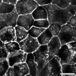
- Western blot analysis of (A) MDCK and (B) Caco-2 cell lysates using Zymed Rb anti-Occludin (C-term-GST) (Product # 42-2400).

- Submitted by
- Invitrogen Antibodies (provider)
- Main image

- Experimental details
- Western blot analysis of (A) MDCK and (B) Caco-2 cell lysates using Zymed Rb anti-Occludin (C-term-GST) (Product # 42-2400).

Supportive validation
- Submitted by
- Invitrogen Antibodies (provider)
- Main image
- Experimental details
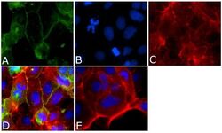
- Indirect immunofluorescent staining of Caco-2 cells using Zymed Rb anti-Occludin (C-term-GST) (Product # 42-2400). Image courtesy of Dr. Jerrold R. Turner, University of Chicago, Chicago, IL.

- Submitted by
- Invitrogen Antibodies (provider)
- Main image
- Experimental details
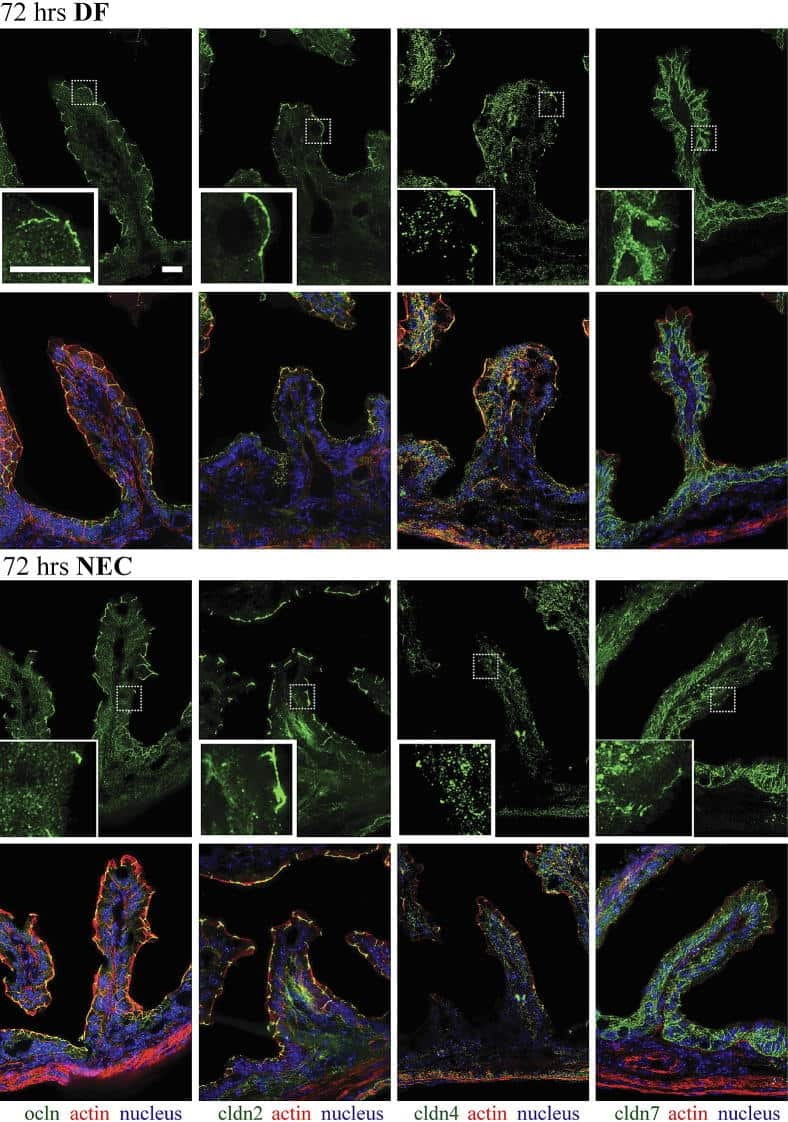
- Immunofluorescent analysis of occludin Antibody was done on 90% confluent log phase CACO2 cells. The cells were fixed with 4% paraformaldehyde for 15 minutes, permeabilized with 0.25% Triton™ X-100 for 10 minutes, and blocked with 5% BSA for 1 hour at room temperature. The cells were labeled with Occludin Antibody (Product # 42-2400) at 1µg/mL in 1% BSA and incubated for 3 hours at room temperature and then labeled with Alexa Fluor 488 Goat Anti-Rabbit IgG Secondary Antibody (Product # A-11008) at a dilution of 1:400 for 45 minutes at room temperature (Panel a: green). Nuclei (Panel b: blue) were stained with SlowFade® Gold Antifade Mountant with DAPI (Product # S36938). F-actin (Panel c: red) was stained with Alexa Fluor 594 Phalloidin (Product # A12381). Panel d is a merged image showing cell junctional localization. Panel e is a no primary antibody control. The images were captured at 40X magnification.

Supportive validation
- Submitted by
- Invitrogen Antibodies (provider)
- Main image

- Experimental details
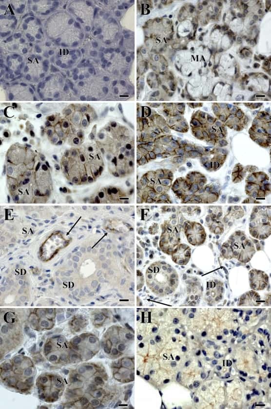
- Immunohistochemistry analysis of Occludin showing staining in the cytoplasm and membrane of paraffin-embedded human kidney tissue (right) compared to a negative control without primary antibody (left). To expose target proteins, antigen retrieval was performed using 10mM sodium citrate (pH 6.0), microwaved for 8-15 min. Following antigen retrieval, tissues were blocked in 3% H2O2-methanol for 15 min at room temperature, washed with ddH2O and PBS, and then probed with a Occludin polyclonal antibody (Product # 42-2400) diluted in 3% BSA-PBS at a dilution of 1:20 overnight at 4ºC in a humidified chamber. Tissues were washed extensively in PBST and detection was performed using an HRP-conjugated secondary antibody followed by colorimetric detection using a DAB kit. Tissues were counterstained with hematoxylin and dehydrated with ethanol and xylene to prep for mounting.

Supportive validation
- Submitted by
- Invitrogen Antibodies (provider)
- Main image
- Experimental details
- NULL
- Submitted by
- Invitrogen Antibodies (provider)
- Main image
- Experimental details
- NULL
- Submitted by
- Invitrogen Antibodies (provider)
- Main image

- Experimental details
- NULL

- Submitted by
- Invitrogen Antibodies (provider)
- Main image

- Experimental details
- NULL

- Submitted by
- Invitrogen Antibodies (provider)
- Main image

- Experimental details
- NULL

- Submitted by
- Invitrogen Antibodies (provider)
- Main image
- Experimental details
- NULL
- Submitted by
- Invitrogen Antibodies (provider)
- Main image

- Experimental details
- Figure 3 MMP13 contributes to tight junction destabilization Source data is available for this figure in the Supporting Information. A-C. Western blot quantification of the protein expression levels of ZO-1 (A), occludin (B) and claudin (C) in mucosal scrapings of ilea from MMP13 +/+ (black) and MMP13 -/- (grey) mice 0 and 8 h after LPS injection ( n = 3-4). D,E. Representative ZO-1 immunogold-labeled TEM images of MMP13 +/+ (D) and MMP13 -/- (E) mice 8 h after LPS injection. F. Schematic overview of the separation of the lipid raft and non-lipid-raft fractions by ultracentrifugation. G. Caveolin-1 western blot analysis of fractions obtained after ultracentrifugation of mucosal scrapings from MMP13 +/+ and MMP13 -/- mice 0 and 8 h after LPS challenge.

- Submitted by
- Invitrogen Antibodies (provider)
- Main image

- Experimental details
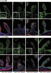
- 4 FIGURE Immunofluorescence staining of Occ and ZO-1 on testis sections. Immunofluorescent staining (arrow) against occludin (Occ) and ZO-1 antibodies (green). DAPI: nucleus (blue). Scale bars indicate 50 mum

- Submitted by
- Invitrogen Antibodies (provider)
- Main image

- Experimental details
- Fig. 1 Infection of Caco-2 cells with C. jejuni disturbs the occludin patterns in a htrA -dependent manner. alpha - Occludin (green) and alpha- C. jejuni immunostaining of Caco-2 mock control cells ( a ) or cells infected with wild-type (wt) strain 81-176 ( b ), Delta htrA knockout mutant ( c ) or Delta htrA complemented with wild-type htrA ( d ). The inlay in panel A shows the belt-like pattern of occludin around the polarized cells as expected (blue arrows). Infection was performed for 12 h at an MOI of 100. The pictures revealed a redistribution of upon wt infection but not Delta htrA mutant (white arrows). Co-localization of occludin with C. jejuni was not observed. DAPI staining (blue) was used for visualization of the DNA in the nuclei
