 Explore
 Validate
 Learn
Learn Western blot
 Immunocytochemistry
Antibody data
- Antibody Data
- Antigen structure
- References [7]
- Comments [0]
- Validations
- Immunocytochemistry [2]
- Other assay [3]
Submit
Validation data
Reference
Comment
Report error
- Product number
- 331594 - Provider product page

- Provider
- Invitrogen Antibodies
- Product name
- Occludin Monoclonal Antibody (OC-3F10), Alexa Fluor™ 594
- Antibody type
- Monoclonal
- Antigen
- Other
- Description
- Reactivity of this antibody with the occludin protein has been confirmed by immunofluorescence and Western blotting (un-conjugated). Tissues/lysates tested: T84 cell line (human intestinal epithelium), MDCK cells (canine kidney), Caco-2 cells (human colon adenocarcinoma), MTE7B (Mouse) and rat liver This antibody reacts specifically with mammalian occludin.
- Reactivity
- Human, Mouse, Rat, Canine
- Host
- Mouse
- Conjugate
- Red dye
- Isotype
- IgG
- Antibody clone number
- OC-3F10
- Vial size
- 100 µg
- Concentration
- 0.5 mg/mL
- Storage
- 4° C, store in dark
Submitted references Dexamethasone Creates a Suppressive Microenvironment and Promotes Aspergillus fumigatus Invasion in a Human 3D Epithelial/Immune Respiratory Model.
The Na+, K+-ATPase β1 subunit regulates epithelial tight junctions via MRCKα.
Three-Dimensional Aggregated Spheroid Model of Hepatocellular Carcinoma Using a 96-Pillar/Well Plate.
Maternal administration of probiotics promotes gut development in mouse offsprings.
Asymmetric Stratification-Induced Polarity Loss and Coordinated Individual Cell Movements Drive Directional Migration of Vertebrate Epithelium.
Occludin, caveolin-1, and Alix form a multi-protein complex and regulate HIV-1 infection of brain pericytes.
Alix-mediated assembly of the actomyosin-tight junction polarity complex preserves epithelial polarity and epithelial barrier.
Luvanda MK, Posch W, Noureen A, Lafon E, Zaderer V, Lass-Flörl C, Wilflingseder D
Journal of fungi (Basel, Switzerland) 2021 Mar 18;7(3)
Journal of fungi (Basel, Switzerland) 2021 Mar 18;7(3)
The Na+, K+-ATPase β1 subunit regulates epithelial tight junctions via MRCKα.
Bai H, Zhou R, Barravecchia M, Norman R, Friedman A, Yu D, Lin X, Young JL, Dean DA
JCI insight 2021 Feb 22;6(4)
JCI insight 2021 Feb 22;6(4)
Three-Dimensional Aggregated Spheroid Model of Hepatocellular Carcinoma Using a 96-Pillar/Well Plate.
Lee SY, Teng Y, Son M, Ku B, Hwang HJ, Tergaonkar V, Chow PK, Lee DW, Nam DH
Molecules (Basel, Switzerland) 2021 Aug 16;26(16)
Molecules (Basel, Switzerland) 2021 Aug 16;26(16)
Maternal administration of probiotics promotes gut development in mouse offsprings.
Yu Y, Lu J, Oliphant K, Gupta N, Claud K, Lu L
PloS one 2020;15(8):e0237182
PloS one 2020;15(8):e0237182
Asymmetric Stratification-Induced Polarity Loss and Coordinated Individual Cell Movements Drive Directional Migration of Vertebrate Epithelium.
Lu Y, Deng R, You H, Xu Y, Antos C, Sun J, Klein OD, Lu P
Cell reports 2020 Oct 13;33(2):108246
Cell reports 2020 Oct 13;33(2):108246
Occludin, caveolin-1, and Alix form a multi-protein complex and regulate HIV-1 infection of brain pericytes.
Torices S, Roberts SA, Park M, Malhotra A, Toborek M
FASEB journal : official publication of the Federation of American Societies for Experimental Biology 2020 Dec;34(12):16319-16332
FASEB journal : official publication of the Federation of American Societies for Experimental Biology 2020 Dec;34(12):16319-16332
Alix-mediated assembly of the actomyosin-tight junction polarity complex preserves epithelial polarity and epithelial barrier.
Campos Y, Qiu X, Gomero E, Wakefield R, Horner L, Brutkowski W, Han YG, Solecki D, Frase S, Bongiovanni A, d'Azzo A
Nature communications 2016 Jun 23;7:11876
Nature communications 2016 Jun 23;7:11876
No comments: Submit comment
Supportive validation
- Submitted by
- Invitrogen Antibodies (provider)
- Main image
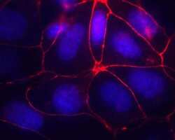
- Experimental details
- Immunofluorescence staining using MTE7B cells with Occludin Monoclonal Antibody, Mouse (OC-3F10), Alexa Fluor® 594 Conjugate: (Product # 331594) DNA is counter-stained with blue Hoechst 33258 (Product # H3569).

- Submitted by
- Invitrogen Antibodies (provider)
- Main image
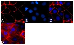
- Experimental details
- Immunofluorescence analysis of Occludin Antibody, Alexa Fluor® 594 conjugate (OC-3F10) was done on 90% confluent log phase CaCo2 cells. The cells were fixed with 4% paraformaldehyde for 15 minutes, permeabilized with 0.25% Triton™ X-100 for 10 minutes, and blocked with 5% BSA for 1 hour at room temperature. The cells were labeled with Occludin Antibody, Alexa Fluor® 594 conjugate (OC-3F10) (Product # 331594) at 1µg/mL in 1% BSA and incubated for 3 hours at room temperature (Panel a: red). Nuclei (Panel b: blue) were stained with SlowFade® Gold Antifade Mountant with DAPI (Product # S36938). Panel c: red). Panel c is a merged image showing cell junctional localization. Panel d is a no primary antibody control. The images were captured at 40X magnification.

Supportive validation
- Submitted by
- Invitrogen Antibodies (provider)
- Main image

- Experimental details
- Figure 3 Western blot analysis of epithelial cell protein and cell proliferation receptor protein expression. Changes in the expression level of the epithelial cell protein ( A , B ), proliferation receptor protein ( C , D ), ECM/ Cell tight junction protein ( E , F ) as determined by western blot analysis. Increased expression of E-cadherin, p-AKT, p-Erk, Fibronectin, ZO-1, and Occludin under ASM culture conditions. All factors were normalized to beta-actin. * p < 0.05, ** p < 0.01, *** p < 0.001, **** p < 0.0001.

- Submitted by
- Invitrogen Antibodies (provider)
- Main image

- Experimental details
- FIGURE 1 HIV-1 infection alters cav-1, ocln, and Alix expression in brain pericytes. Pericytes were either mock-infected or infected with 60 ng/mL HIV-1 p24 for 48 h (A) or 72 h (B). The expression of cav-1, ocln, and Alix was evaluated by immunoblotting. GAPDH was used as a loading control. C, Representative immunostaining images of cav-1 (green), ocln (red), and Alix (purple) in mock-infected or HIV-1-infected for 48 or 72 h. Alix expression intensity was increased by the same factors in all groups to allow for better visualization. Graphs show the mean +- SD from three independent experiments. **** P < .0001, ** P = .003, * P < .0449, n = 4-9 per group; scale bars, 20 um

- Submitted by
- Invitrogen Antibodies (provider)
- Main image

- Experimental details
- FIGURE 2 Cav-1, ocln, and Alix form a stable complex in mock-infected and HIV-1-infected pericytes. A, Diagram illustrating Alix and its binding partners identified in this study in a structural ribbon representation. The ESCRT-associated protein Alix binds tyrosine motifs via its C-terminal Proline Rich Domain (PRD). Caveolae are made up of oligomers of cav-1 and cavin proteins, and the figure shows a trimeric coiled coil for Cavin4a HR1 domain. For ocln, the region shown is the cytoplasmic C-terminal region that is known to bind scaffolding proteins. Figure was prepared using PyMol with the listed PDB accession numbers. B-E, Pericytes were either mock-infected (B) or infected with HIV-1 (C) for 48 h as in Figure 1 . A total of 600 ug of cell lysate protein was incubated with cav-1 antibody for 24 h, followed by incubation with protein A/G PLUS-Agarose beads. Both immunoprecipitates and supernatants were analyzed by immunoblotting for the presence of cav-1, ocln, and Alix. D, Immunostaining of cav-1 (green), ocln (red), and Alix (purple) in mock-infected or HIV-1-infected for 48 h. E, Cellular localization of the cav-1, ocln, and Alix complex in mock-infected or HIV-1-infected for 48 h. Cell membranes are visualized by blue fluorescent dye DiB (left panel). Middle panel, the cav-1, ocln, and Alix complexes merged with a bright field image of a single pericyte. The images were analyzed as in Figure 1 ; n = 4 per group; scale bar 5, 10, or 20 um
