 Explore
 Validate
 Learn
Learn Western blot
Antibody data
- Antibody Data
- Antigen structure
- References [5]
- Comments [0]
- Validations
- Western blot [2]
- Immunocytochemistry [2]
- Immunohistochemistry [3]
- Other assay [3]
Submit
Validation data
Reference
Comment
Report error
- Product number
- 710192 - Provider product page

- Provider
- Invitrogen Antibodies
- Product name
- Occludin Recombinant Polyclonal Antibody (6HCLC)
- Antibody type
- Polyclonal
- Antigen
- Synthetic peptide
- Description
- Recombinant rabbit polyclonal antibodies are unique offerings from Thermo Fisher Scientific. They are comprised of a selection of multiple different recombinant monoclonal antibodies, providing the best of both worlds - the sensitivity of polyclonal antibodies with the specificity of monoclonal antibodies - all delivered with the consistency only found in a recombinant antibody. While functionally the same as a polyclonal antibody - recognizing multiple epitope sites on the target and producing higher detection sensitivity for low abundance targets - a recombinant rabbit polyclonal antibody has a known mixture of light and heavy chains. The exact population can be produced in every lot, circumventing the biological variability typically associated with polyclonal antibody production.
- Reactivity
- Human, Mouse
- Host
- Rabbit
- Isotype
- IgG
- Antibody clone number
- 6HCLC
- Vial size
- 100 µg
- Concentration
- 0.5 mg/mL
- Storage
- Store at 4°C short term. For long term storage, store at -20°C, avoiding freeze/thaw cycles.
Submitted references TIMP1 preserves the blood-brain barrier through interacting with CD63/integrin β 1 complex and regulating downstream FAK/RhoA signaling.
TatS: a novel in vitro tattooed human skin model for improved pigment toxicology research.
Protein Kinase D2 Protects against Acute Colitis Induced by Dextran Sulfate Sodium in Mice.
α-Synuclein pre-formed fibrils impair tight junction protein expression without affecting cerebral endothelial cell function.
Modified Pulsatilla decoction attenuates oxazolone-induced colitis in mice through suppression of inflammation and epithelial barrier disruption.
Tang J, Kang Y, Huang L, Wu L, Peng Y
Acta pharmaceutica Sinica. B 2020 Jun;10(6):987-1003
Acta pharmaceutica Sinica. B 2020 Jun;10(6):987-1003
TatS: a novel in vitro tattooed human skin model for improved pigment toxicology research.
Hering H, Zoschke C, Kühn M, Gadicherla AK, Weindl G, Luch A, Schreiver I
Archives of toxicology 2020 Jul;94(7):2423-2434
Archives of toxicology 2020 Jul;94(7):2423-2434
Protein Kinase D2 Protects against Acute Colitis Induced by Dextran Sulfate Sodium in Mice.
Xiong J, Zhou MF, Wang YD, Chen LP, Xu WF, Wang YD, Deng F, Liu SD
Scientific reports 2016 Sep 23;6:34079
Scientific reports 2016 Sep 23;6:34079
α-Synuclein pre-formed fibrils impair tight junction protein expression without affecting cerebral endothelial cell function.
Kuan WL, Bennett N, He X, Skepper JN, Martynyuk N, Wijeyekoon R, Moghe PV, Williams-Gray CH, Barker RA
Experimental neurology 2016 Nov;285(Pt A):72-81
Experimental neurology 2016 Nov;285(Pt A):72-81
Modified Pulsatilla decoction attenuates oxazolone-induced colitis in mice through suppression of inflammation and epithelial barrier disruption.
Wang X, Fan F, Cao Q
Molecular medicine reports 2016 Aug;14(2):1173-9
Molecular medicine reports 2016 Aug;14(2):1173-9
No comments: Submit comment
Supportive validation
- Submitted by
- Invitrogen Antibodies (provider)
- Main image
- Experimental details
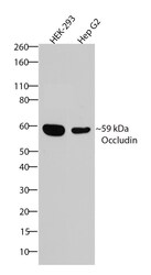
- Western blot analysis of Occludin was performed by loading 30 µg of HEK-293 and Hep G2 cell lysates using Novex®NuPAGE®4-12% Bis-Tris gel (Product # NP0321BOX), XCell SureLock Electrophoresis System (Product # EI0002), Novex® Sharp Pre-Stained Protein Standard (Product # LC5800), and iBlot® Dry Blotting System (Product # IB21001). Proteins were transferred to a nitrocellulose membrane and blocked with 5% skim milk for 1 hour at room temperature. Occludin was detected at ~59 kDa using Occludin Recombinant Rabbit Polyclonal Antibody (Product # 710192) at a 1:1000 dilution in 2.5% skim milk at 4°C overnight on a rocking platform. Detection was performed using an HRP-conjugated Goat anti-Rabbit secondary antibody (Product # G-21234) at a 1:5000 dilution and chemiluminescent detection was performed using Pierce™ ECL Western blotting Substrate (Product # 32106).
- Submitted by
- Invitrogen Antibodies (provider)
- Main image
- Experimental details
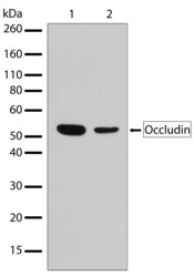
- Western blot analysis of Occludin in whole cell extracts of HEK (lane 1) and HepG2 (lane 2) using an Occludin Recombinant Rabbit Polyclonal Antibody (Product # 710192) at a dilution of 2 µg/mL. Samples were detected using chemiluminescence (ECL). Results show a band at ~59kDa.

Supportive validation
- Submitted by
- Invitrogen Antibodies (provider)
- Main image

- Experimental details
- Immunofluorescent analysis of Occludin in HEK293 cells using an Occludin Recombinant Rabbit Polyclonal Antibody (Product # 710192) followed by detection using an Alexa Fluor 488-conjugated Goat anti-Rabbit secondary antibody (green) and nuclei stained using DAPI (blue) (image A). Image B is a composite image showing localization of Occludin at tight junctions.

- Submitted by
- Invitrogen Antibodies (provider)
- Main image

- Experimental details
- Immunofluorescent analysis of Occludin was performed on 90% confluent log phase Caco-2 cells. The cells were fixed with 4% paraformaldehyde for 15 minutes, and blocked with 5% BSA for 1 hour at room temperature. The cells were labeled with Occludin Recombinant Rabbit Polyclonal Antibody (Product # 710192) at a dilution of 1:500 in 1% BSA and incubated for 3 hours at room temperature and then labeled with Alexa Fluor® 488 Goat anti-Rabbit IgG secondary antibody (Product # A-11008) at a dilution of 1:400 for 30 minutes at room temperature (Panel a: green). Nuclei (Panel b: blue) were stained with SlowFade® Gold Antifade Mountant with DAPI (Product # S36938). Panel c is a merged image showing cell junction localization and panel d is a control without primary antibody. The images were captured using a Nikon microscope at 20X magnification.

Supportive validation
- Submitted by
- Invitrogen Antibodies (provider)
- Main image
- Experimental details
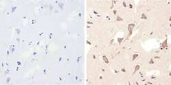
- Immunohistochemistry analysis of Occludin showing staining in the cytoplasm of paraffin-embedded human brain tissue (right) compared to a negative control without primary antibody (left). To expose target proteins, antigen retrieval was performed using 10 mM sodium citrate (pH 6.0), microwaved for 8-15 min. Following antigen retrieval, tissues were blocked in 3% H2O2-methanol for 15 min at room temperature, washed with ddH2O and PBS, and then probed with a Occludin (6H10L9) Recombinant Rabbit Polyclonal Antibody (Product # 710192) diluted in 3% BSA-PBS at a dilution of 1:20 overnight at 4°C in a humidified chamber. Tissues were washed extensively in PBST and detection was performed using a HRP-conjugated secondary antibody followed by colorimetric detection using a DAB kit. Tissues were counterstained with hematoxylin and dehydrated with ethanol and xylene to prep for mounting.

- Submitted by
- Invitrogen Antibodies (provider)
- Main image

- Experimental details
- Immunohistochemistry analysis of Occludin showing staining in the cytoplasm of paraffin-embedded human kidney tissue (right) compared to a negative control without primary antibody (left). To expose target proteins, antigen retrieval was performed using 10 mM sodium citrate (pH 6.0), microwaved for 8-15 min. Following antigen retrieval, tissues were blocked in 3% H2O2-methanol for 15 min at room temperature, washed with ddH2O and PBS, and then probed with Occludin (6H10L9) Recombinant Rabbit Polyclonal Antibody (Product # 710192) diluted in 3% BSA-PBS at a dilution of 1:20 overnight at 4°C in a humidified chamber. Tissues were washed extensively in PBST and detection was performed using a HRP-conjugated secondary antibody followed by colorimetric detection using a DAB kit. Tissues were counterstained with hematoxylin and dehydrated with ethanol and xylene to prep for mounting.

- Submitted by
- Invitrogen Antibodies (provider)
- Main image

- Experimental details
- Immunohistochemistry analysis of Occludin showing staining in the cytoplasm of paraffin-embedded mouse kidney tissue (right) compared to a negative control without primary antibody (left). To expose target proteins, antigen retrieval was performed using 10 mM sodium citrate (pH 6.0), microwaved for 8-15 min. Following antigen retrieval, tissues were blocked in 3% H2O2-methanol for 15 min at room temperature, washed with ddH2O and PBS, and then probed with a Occludin (6H10L9) Recombinant Rabbit Polyclonal Antibody (Product # 710192) diluted in 3% BSA-PBS at a dilution of 1:20 overnight at 4°C in a humidified chamber. Tissues were washed extensively in PBST and detection was performed using a HRP-conjugated secondary antibody followed by colorimetric detection using a DAB kit. Tissues were counterstained with hematoxylin and dehydrated with ethanol and xylene to prep for mounting.

Supportive validation
- Submitted by
- Invitrogen Antibodies (provider)
- Main image

- Experimental details
- Fig. 2 Histological characterization of tattooed human skin models (TatS). The columns show (from top to bottom): haematoxylin and eosin (H&E) stain and immunofluorescence staining of occludin-1, filaggrin, E-cadherin, collagen IV and tenascin-C (all in red). Immunofluorescence pictures were counterstained with Hoechst 33258 (blue), indicating the cell nuclei. Dotted lines in H&E pictures indicate the pigment border. Black bar for H&E stain equals 100 um, white bars equal 50 um. w/o, without; titanium dioxide, TiO 2 ; Pigment Orange 13, P.O.13

- Submitted by
- Invitrogen Antibodies (provider)
- Main image
- Experimental details
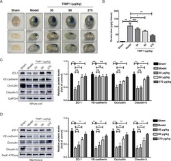
- Figure 2 rTIMP1 treatment attenuates blood-brain barrier (BBB) permeability and loss of junctional proteins (JPs) in TBI mice. (A) Representative images of brain tissues and corresponding coronal sections from sham injury group, TBI group treated with vehicle or TBI groups treated with rTIMP1 (30, 90, and 270 mug/kg, i.v.) at 3 days after traumatic brain injury. Blue area indicates extravasation of Evans blue dye. (B) Quantification of the Evans blue dye contents leaking into the ipsilateral cerebral hemisphere tissue of mice from the indicated treatment groups ( n = 11-12 per group). (C) Western blot analysis and quantification of ZO-1, VE-cadherin, occludin and claudin-5 in ipsilateral hemispheric brain total lysates from the indicated treatment groups at 72 h post-TBI. GAPDH was used as loading control. n = 5 per group. (D) Western blot analysis and quantification of ZO-1, VE-cadherin, occludin and claudin-5 in ipsilateral hemispheric brain membrane fragments from the indicated treatment groups at 72 h post-TBI. Na/K ATPase was used as loading control. Data are presented as the mean +- SEM, n = 5 per group. (ns, not significant; * P < 0.05; ** P < 0.01). Figure 2

- Submitted by
- Invitrogen Antibodies (provider)
- Main image

- Experimental details
- Figure 4 TIMP1 attenuates BBB impairment induced by hypoxia plus IL-1 beta insult. HBMECs treated with PBS or rTIMP1 at indicated doses were subjected to hypoxia plus 20 ng/mL IL-1 beta for 24 h. (A) HBMECs from the indicated treatment groups were subject to transwell permeability assay. Data represent mean +- SEM of six independent experiments (ns, not significant; ** P < 0.01). (B) HBMECs from the indicated treatment groups were subject to TEER assay. Data represent mean +- SEM of six independent experiments (ns, not significant; ** P < 0.01). (C) Western blot analysis and quantification of ZO-1, occludin, claudin-5 and VE-cadherin protein levels in total cell lysates from the indicated treatment groups. GAPDH was used as loading control. Data represent mean +- SEM of three independent experiments (ns, not significant; * P < 0.05; ** P < 0.01). (D) Western blot analysis and quantification of ZO-1, occludin, claudin-5 and VE-cadherin protein levels on membrane fragments from the indicated treatment groups. Na/K ATPase was used as loading control. Data represent mean +- SEM of three independent experiments (ns, not significant; * P < 0.05; ** P < 0.01). Figure 4
