Explore
 Validate
 Learn
Learn Western blot
 Immunocytochemistry
Antibody data
- Antibody Data
- Antigen structure
- References [1]
- Comments [0]
- Validations
- Immunocytochemistry [1]
Submit
Validation data
Reference
Comment
Report error
- Product number
- HPA001813 - Provider product page

- Provider
- Atlas Antibodies
- Proper citation
- Atlas Antibodies Cat#HPA001813, RRID:AB_1080764
- Product name
- Anti-TJP2
- Antibody type
- Polyclonal
- Description
- Polyclonal Antibody against Human TJP2, Gene description: tight junction protein 2, Alternative Gene Names: DFNA51, X104, ZO-2, ZO2, Validated applications: ICC, IHC, WB, Uniprot ID: Q9UDY2, Storage: Store at +4°C for short term storage. Long time storage is recommended at -20°C.
- Reactivity
- Human
- Host
- Rabbit
- Conjugate
- Unconjugated
- Isotype
- IgG
- Vial size
- 100 µl
- Concentration
- 0.2 mg/ml
- Storage
- Store at +4°C for short term storage. Long time storage is recommended at -20°C.
- Handling
- The antibody solution should be gently mixed before use.
Submitted references Sequential development of intercellular junctions in bioengineered human corneas
González-Andrades M, Garzón I, Gascón M, Muñoz-Ávila J, Sánchez-Quevedo M, Campos A, Alaminos M
Journal of Tissue Engineering and Regenerative Medicine 2009;3(6):442-449
Journal of Tissue Engineering and Regenerative Medicine 2009;3(6):442-449
No comments: Submit comment
Supportive validation
- Submitted by
- Atlas Antibodies (provider)
- Main image
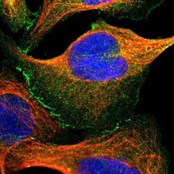
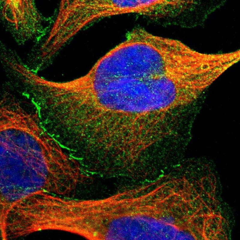
- Experimental details
- Immunofluorescent staining of human cell line U-2 OS shows positivity in plasma membrane, cytoplasm & cell junctions.
- Sample type
- Human