 Explore
 Validate
 Learn
Learn Western blot
Antibody data
- Antibody Data
- Antigen structure
- References [4]
- Comments [0]
- Validations
- Western blot [8]
- Immunocytochemistry [1]
- Immunohistochemistry [1]
- Other assay [1]
Submit
Validation data
Reference
Comment
Report error
- Product number
- PA5-30604 - Provider product page

- Provider
- Invitrogen Antibodies
- Product name
- SOD2 Polyclonal Antibody
- Antibody type
- Polyclonal
- Antigen
- Recombinant full-length protein
- Description
- Recommended positive controls: HeLa, HCT116, mouse brain, rat brain, 293T.
- Concentration
- 1 mg/mL
Submitted references Preventive Effect of Limosilactobacillus fermentum SCHY34 on Lead Acetate-Induced Neurological Damage in SD Rats.
Pre-treatment With PLGA/Silibinin Nanoparticles Mitigates Dacarbazine-Induced Hepatotoxicity.
Oxidative stress and mitochondrial uncoupling protein 2 expression in hepatic steatosis induced by exposure to xenobiotic DDE and high fat diet in male Wistar rats.
Extracts of Physalis peruviana Protect Astrocytic Cells Under Oxidative Stress With Rotenone.
Long X, Wu H, Zhou Y, Wan Y, Kan X, Gong J, Zhao X
Frontiers in nutrition 2022;9:852012
Frontiers in nutrition 2022;9:852012
Pre-treatment With PLGA/Silibinin Nanoparticles Mitigates Dacarbazine-Induced Hepatotoxicity.
Durymanov M, Permyakova A, Reineke J
Frontiers in bioengineering and biotechnology 2020;8:495
Frontiers in bioengineering and biotechnology 2020;8:495
Oxidative stress and mitochondrial uncoupling protein 2 expression in hepatic steatosis induced by exposure to xenobiotic DDE and high fat diet in male Wistar rats.
Migliaccio V, Scudiero R, Sica R, Lionetti L, Putti R
PloS one 2019;14(4):e0215955
PloS one 2019;14(4):e0215955
Extracts of Physalis peruviana Protect Astrocytic Cells Under Oxidative Stress With Rotenone.
Areiza-Mazo N, Robles J, Zamudio-Rodriguez JA, Giraldez L, Echeverria V, Barrera-Bailon B, Aliev G, Sahebkar A, Ashraf GM, Barreto GE
Frontiers in chemistry 2018;6:276
Frontiers in chemistry 2018;6:276
No comments: Submit comment
Supportive validation
- Submitted by
- Invitrogen Antibodies (provider)
- Main image

- Experimental details
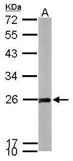
- Western blot analysis of SOD2 using 30 µg of A) HeLa and B) HCT116 lysate. Samples were loaded onto a 15% SDS-PAGE gel and probed with a SOD2 polyclonal antibody (Product # PA5-30604) at a dilution of 1:1000.

- Submitted by
- Invitrogen Antibodies (provider)
- Main image

- Experimental details
- Western blot analysis of SOD2 using 50 µg of mouse brain lysate. Samples were loaded onto a 12% SDS-PAGE gel and probed with a SOD2 polyclonal antibody (Product # PA5-30604) at a dilution of 1:1000.
- Submitted by
- Invitrogen Antibodies (provider)
- Main image

- Experimental details
- Western blot analysis of SOD2 using 50 µg of rat brain lysate. Samples were loaded onto a 12% SDS-PAGE gel and probed with a SOD2 polyclonal antibody (Product # PA5-30604) at a dilution of 1:1000.

- Submitted by
- Invitrogen Antibodies (provider)
- Main image

- Experimental details
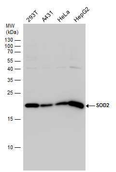
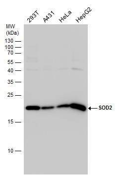
- Western Blot analysis of SOD2 was performed by separating 30 µg of various whole cell extracts by 15% SDS-PAGE. Proteins were transferred to a membrane and probed with a SOD2 Polyclonal Antibody (Product # PA5-30604) at a dilution of 1:1000 and a HRP-conjugated anti-rabbit IgG secondary antibody.
- Submitted by
- Invitrogen Antibodies (provider)
- Main image

- Experimental details
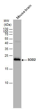
- Western Blot analysis of SOD2 was performed by separating 50 µg of mouse tissue extracts by 15% SDS-PAGE. Proteins were transferred to a membrane and probed with a SOD2 Polyclonal Antibody (Product # PA5-30604) at a dilution of 1:1000. The HRP-conjugated anti-rabbit IgG antibody was used to detect the primary antibody.
- Submitted by
- Invitrogen Antibodies (provider)
- Main image

- Experimental details
- Western Blot analysis of SOD2 was performed by separating 50 µg of rat tissue extracts by 15% SDS-PAGE. Proteins were transferred to a membrane and probed with a SOD2 Polyclonal Antibody (Product # PA5-30604) at a dilution of 1:2500. The HRP-conjugated anti-rabbit IgG antibody was used to detect the primary antibody.

- Submitted by
- Invitrogen Antibodies (provider)
- Main image
- Experimental details
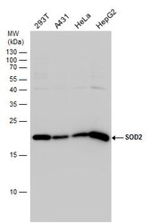
- Western Blot analysis of SOD2 was performed by separating 30 µg of various whole cell extracts by 15% SDS-PAGE. Proteins were transferred to a membrane and probed with a SOD2 Polyclonal Antibody (Product # PA5-30604) at a dilution of 1:1000 and a HRP-conjugated anti-rabbit IgG secondary antibody.
- Submitted by
- Invitrogen Antibodies (provider)
- Main image

- Experimental details
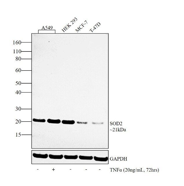
- Western blot analysis was performed on whole cell extracts of A549 (Lane 1), A549 treated with TNFa (20 ng/mL, 72hrs) (Lane 2), HEK 293 (Lane 3), MCF-7 (Lane 4) and T-47D (Lane 5). The blot was probed with SOD2 Polyclonal Antibody (Product # PA5-30604, 1:1000 dilution) and detected by chemiluminescence using Goat anti-Rabbit IgG (H+L) Superclonal™ Secondary Antibody, HRP conjugate (Product # A27036, 0.25 µg/mL, 1:4000 dilution). A 21 kDa band corresponding to SOD2 was observed in all the lanes, with A549 showing an increase in expression upon TNFa treatment.
Supportive validation
- Submitted by
- Invitrogen Antibodies (provider)
- Main image

- Experimental details
- Immunofluorescent analysis of SOD2 in methanol-fixed HeLa cells using a SOD2 polyclonal antibody (Product # PA5-30604) at a 1:200 dilution.

Supportive validation
- Submitted by
- Invitrogen Antibodies (provider)
- Main image

- Experimental details
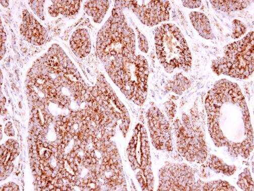
- Immunohistochemistry (Paraffin) analysis of SOD2 was performed in paraffin-embedded human breast carcinoma tissue using SOD2 Polyclonal Antibody (Product # PA5-30604) at a dilution of 1:500.
Supportive validation
- Submitted by
- Invitrogen Antibodies (provider)
- Main image

- Experimental details
- Figure 4 Expression of antioxidant defense and ERK proteins in T98G cells. (A) Expression of Superoxide dismutase (SOD) and Catalase determined by Western Blot in T98G cell cultures after co-treatment with 50 muM rotenone plus EF and AD at 25 mu g/ml. (B) Expression of Glutathione determined by Western Blot in T98G cell cultures after co-treatment with 50 muM rotenone plus EF and AD at 25 mu g/ml. (C) Expression of ERK and MAPK proteins determined by Western blotting on T98G cells after co-treatment with rotenone plus EF and AD at 25 mu g/ml.
