 Explore
 Validate
 Learn
Learn Western blot
 Immunocytochemistry
Antibody data
- Antibody Data
- Antigen structure
- References [4]
- Comments [0]
- Validations
- Western blot [7]
- Immunocytochemistry [1]
- Immunohistochemistry [3]
Submit
Validation data
Reference
Comment
Report error
- Product number
- GTX100659 - Provider product page

- Provider
- GeneTex
- Proper citation
- GeneTex Cat#GTX100659, RRID:AB_1951972
- Product name
- SOD1 antibody
- Antibody type
- Polyclonal
- Reactivity
- Human, Mouse, Rat
- Host
- Rabbit
Submitted references Reduced camptothecin sensitivity of estrogen receptor-positive human breast cancer cells following exposure to di(2-ethylhexyl)phthalate (DEHP) is associated with DNA methylation changes.
Exogenous C₈-Ceramide Induces Apoptosis by Overproduction of ROS and the Switch of Superoxide Dismutases SOD1 to SOD2 in Human Lung Cancer Cells.
Bioavailability of andrographolide and protection against carbon tetrachloride-induced oxidative damage in rats.
Altered expression of atypical PKC and Ryk in the spinal cord of a mouse model of amyotrophic lateral sclerosis.
Chou CK, Huang HW, Yang CF, Dahms HU, Liang SS, Wang TN, Kuo PL, Hsi E, Tsai EM, Chiu CC
Environmental toxicology 2019 Apr;34(4):401-414
Environmental toxicology 2019 Apr;34(4):401-414
Exogenous C₈-Ceramide Induces Apoptosis by Overproduction of ROS and the Switch of Superoxide Dismutases SOD1 to SOD2 in Human Lung Cancer Cells.
Chang YC, Fong Y, Tsai EM, Chang YG, Chou HL, Wu CY, Teng YN, Liu TC, Yuan SS, Chiu CC
International journal of molecular sciences 2018 Oct 2;19(10)
International journal of molecular sciences 2018 Oct 2;19(10)
Bioavailability of andrographolide and protection against carbon tetrachloride-induced oxidative damage in rats.
Chen HW, Huang CS, Li CC, Lin AH, Huang YJ, Wang TS, Yao HT, Lii CK
Toxicology and applied pharmacology 2014 Oct 1;280(1):1-9
Toxicology and applied pharmacology 2014 Oct 1;280(1):1-9
Altered expression of atypical PKC and Ryk in the spinal cord of a mouse model of amyotrophic lateral sclerosis.
Tury A, Tolentino K, Zou Y
Developmental neurobiology 2014 Aug;74(8):839-50
Developmental neurobiology 2014 Aug;74(8):839-50
No comments: Submit comment
Enhanced validation
Supportive validation
- Submitted by
- GeneTex (provider)
- Enhanced method
- Genetic validation
- Main image
- Experimental details
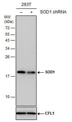
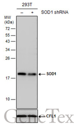
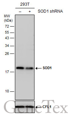
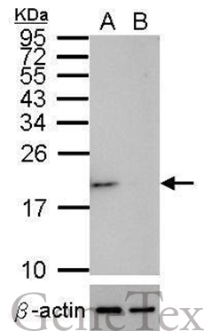
- Non-transfected (¡V) and transfected (+) 293T whole cell extracts (30 ?g) were separated by 15% SDS-PAGE, and the membrane was blotted with SOD1 antibody (GTX100659) diluted at 1:5000. The HRP-conjugated anti-rabbit IgG antibody (GTX213110-01) was used to detect the primary antibody.

Supportive validation
- Submitted by
- GeneTex (provider)
- Main image
- Experimental details
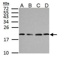
- SOD1 antibody detects SOD1 protein by western blot analysis.A.30 ?g NT2D1 whole cell lysate/extract B.30 ?g PC-3 whole cell lysate/extract C.30 ?g U87-MG whole cell lysate/extractD.30 ?g SK-N-SH whole cell lysate/extract15% SDS-PAGESOD1 antibody (GTX100659) dilution: 1:500 The HRP-conjugated anti-rabbit IgG antibody (GTX213110-01) was used to detect the primary antibody.
- Submitted by
- GeneTex (provider)
- Main image
- Experimental details
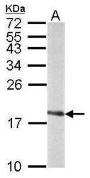
- Sample (50 ?g of whole cell lysate) A: Rat brain 15% SDS PAGE GTX100659 diluted at 1:1000 The HRP-conjugated anti-rabbit IgG antibody (GTX213110-01) was used to detect the primary antibody.

- Submitted by
- GeneTex (provider)
- Main image

- Experimental details
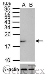
- Western blot analysis of SOD1 (GTX100659, upper panel) and beta-actin (GTX109639, lower panel)? Sample (30 ?g of whole cell lysate)? A: HeLa mock control? B: HeLa transfected shSOD1 15% SDS PAGE? GTX100659 diluted at 1:500 The HRP-conjugated anti-rabbit IgG antibody (GTX213110-01) was used to detect the primary antibody.

- Submitted by
- GeneTex (provider)
- Main image
- Experimental details
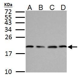
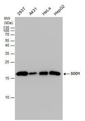
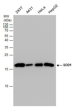
- SOD1 antibody detects SOD1 protein by western blot analysis. Various whole cell extracts (30 ?g) were separated by 15% SDS-PAGE, and the membrane was blotted with SOD1 antibody (GTX100659) diluted by 1:1000. The HRP-conjugated anti-rabbit IgG antibody (GTX213110-01) was used to detect the primary antibody.
- Submitted by
- GeneTex (provider)
- Main image
- Experimental details
- Non-transfected (¡V) and transfected (+) 293T whole cell extracts (30 ?g) were separated by 15% SDS-PAGE, and the membrane was blotted with SOD1 antibody (GTX100659) diluted at 1:5000. The HRP-conjugated anti-rabbit IgG antibody (GTX213110-01) was used to detect the primary antibody.
- Submitted by
- GeneTex (provider)
- Main image
- Experimental details
- Western blot analysis of SOD1 (GTX100659, upper panel) and beta-actin (GTX109639, lower panel)? Sample (30 ?g of whole cell lysate)? A: HeLa mock control? B: HeLa transfected shSOD1 15% SDS PAGE? GTX100659 diluted at 1:500 The HRP-conjugated anti-rabbit IgG antibody (GTX213110-01) was used to detect the primary antibody.
Supportive validation
- Submitted by
- GeneTex (provider)
- Main image
- Experimental details
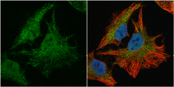
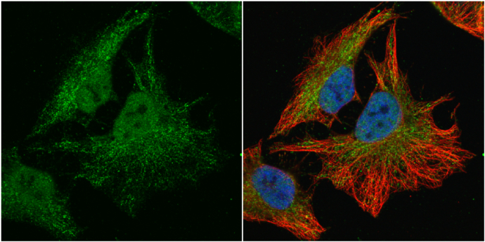
- SOD1 antibody detects SOD1 protein at cytoplasm and nucleus by immunofluorescent analysis.Sample: HeLa cells were fixed in 4% paraformaldehyde at RT for 15 min.Green: SOD1 protein stained by SOD1 antibody (GTX100659) diluted at 1:100.Red: alpha Tubulin, a cytoskeleton marker, stained by alpha Tubulin antibody [GT114] (GTX628802) diluted at 1:1000.Blue: Hoechst 33342 staining.
Supportive validation
- Submitted by
- GeneTex (provider)
- Main image

- Experimental details
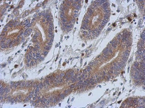
- Immunohistochemical analysis of paraffin-embedded human colon carcinoma, using SOD1(GTX100659) antibody at 1:500 dilution.
- Submitted by
- GeneTex (provider)
- Main image
- Experimental details
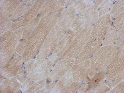
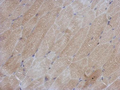
- Immunohistochemical analysis of paraffin-embedded mouse muscle, using SOD1(GTX100659) antibody at 1:500 dilution.
- Submitted by
- GeneTex (provider)
- Main image
- Experimental details
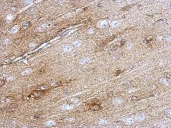
- Immunohistochemical analysis of paraffin-embedded rat brain, using SOD1(GTX100659) antibody at 1:500 dilution.
