Explore
 Validate
 Learn
Learn Western blot
 ELISA
ELISAAntibody data
- Antibody Data
- Antigen structure
- References [1]
- Comments [0]
- Validations
- ELISA [3]
- Immunocytochemistry [1]
- Immunohistochemistry [4]
- Other assay [2]
Submit
Validation data
Reference
Comment
Report error
- Product number
- PA5-85095 - Provider product page

- Provider
- Invitrogen Antibodies
- Product name
- SOD1 Polyclonal Antibody
- Antibody type
- Polyclonal
- Antigen
- Recombinant full-length protein
- Description
- Antigen Retrieval (IF): citrate buffer, pH 6.0, 15 min, and IHC: EDTA based, pH 8.0 buffer, 15min. Keep as concentrated solution. Predicted reactivity: Mouse (81%), Rat (81%), Pig (80%), Rabbit (82%), Rhesus Monkey (91%), Chimpanzee (100%). Positive Control: 293T, HeLa, NT2D1, SK-N-AS, IMR32, SK-N-SH, U87-MG, mouse brain, Rat brain, F79, F123. Store product as a concentrated solution. Centrifuge briefly prior to opening the vial.
- Reactivity
- Human, Mouse, Rat, Chicken/Avian
- Host
- Rabbit
- Isotype
- IgG
- Vial size
- 100 μL
- Concentration
- 0.15 mg/mL
- Storage
- Store at 4°C short term. For long term storage, store at -20°C, avoiding freeze/thaw cycles.
Submitted references β-Nicotinamide Mononucleotide (NMN) Administrated by Intraperitoneal Injection Mediates Protection Against UVB-Induced Skin Damage in Mice.
Zhou X, Du HH, Long X, Pan Y, Hu J, Yu J, Zhao X
Journal of inflammation research 2021;14:5165-5182
Journal of inflammation research 2021;14:5165-5182
No comments: Submit comment
Supportive validation
- Submitted by
- Invitrogen Antibodies (provider)
- Main image
- Experimental details
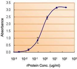
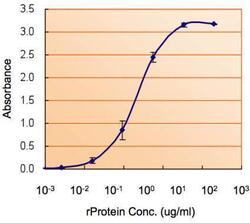
- ELISA detection of SOD1 using SOD1 Polyclonal Antibody (Product # PA5-85095) for capture at a concentration of 5 µg/mL and a SOD1 Polyclonal Antibody for detection at a concentration of 1.5 µg/mL.

- Submitted by
- Invitrogen Antibodies (provider)
- Main image
- Experimental details
- ELISA detection of SOD1 using SOD1 Polyclonal Antibody (Product # PA5-85095) for capture at a concentration of 5 µg/mL and a SOD1 Polyclonal Antibody for detection at a concentration of 1.5 µg/mL.

- Submitted by
- Invitrogen Antibodies (provider)
- Main image

- Experimental details
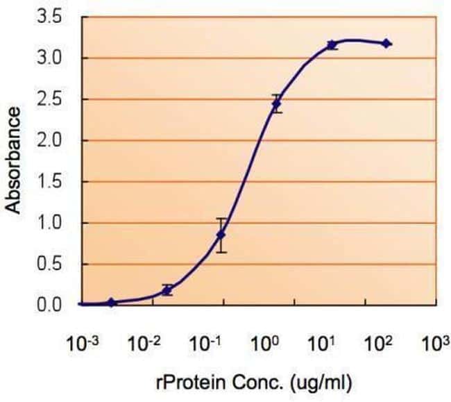
- ELISA analysis of SOD1 using SOD1 polyclonal antibody (Product # PA5-85095) at a dilution of 1.5 µg/mL.

Supportive validation
- Submitted by
- Invitrogen Antibodies (provider)
- Main image
- Experimental details
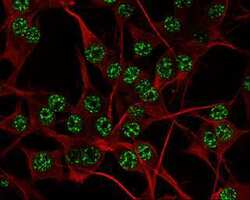
- SOD1 Polyclonal Antibody detects SOD1 protein at nucleus by immunofluorescent analysis. Sample: U-87 MG cells were fixed in 4% paraformaldehyde at RT for 15 min. Green: SOD1 protein stained by SOD1 Polyclonal Antibody (Product # PA5-85095) diluted at 1:500. Red: beta Tubulin 3/ TUJ1 protein stained by beta-3 Tubulin Polyclonal Antibody diluted at 1:200.

Supportive validation
- Submitted by
- Invitrogen Antibodies (provider)
- Main image

- Experimental details
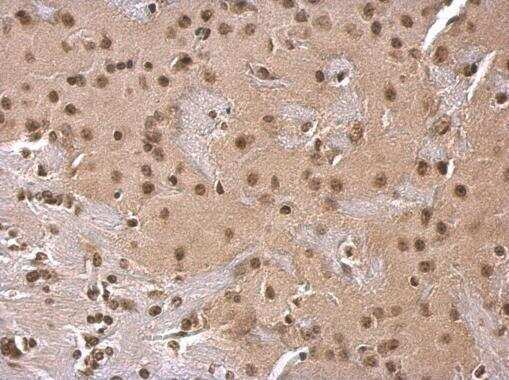
- SOD1 Polyclonal Antibody detects SOD1 protein at cytosol on mouse fore brain by immunohistochemical analysis. Sample: Paraffin-embedded mouse fore brain. SOD1 Polyclonal Antibody (Product # PA5-85095) dilution: 1:500. Antigen Retrieval: EDTA based buffer, pH 8.0, 15 min.
- Submitted by
- Invitrogen Antibodies (provider)
- Main image
- Experimental details
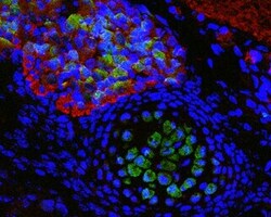
- Immunohistochemistry (Paraffin) photomicrographs of paraffin-embedded mouse fetal brain. Green: SOD1 Polyclonal Antibody (Product # PA5-85095) diluted at 1:200. The signal was developed using goat anti-rabbit IgG antibody (Dylight488). Red: beta Tubulin 3/ TUJ1 antibody [GT11710] diluted at 1:100. The signal was developed using goat anti-mouse IgG antibody (Dylight594). Blue: Nuclear staining with Hoechst 33342. Antigen Retrieval: Citrate buffer, pH 6.0, 15 min.

- Submitted by
- Invitrogen Antibodies (provider)
- Main image

- Experimental details
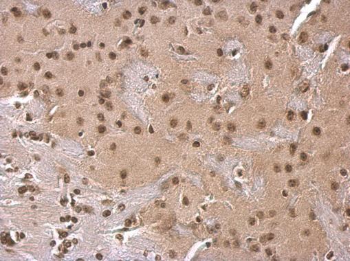
- Immunohistochemistry analysis of SOD1 in paraffin-embedded mouse fore brain using SOD1 polyclonal antibody (Product # PA5-85095) at a dilution of 1:500.
- Submitted by
- Invitrogen Antibodies (provider)
- Main image

- Experimental details
- Immunohistochemistry analysis of SOD1 in paraffin-embedded mouse fetal brain using SOD1 polyclonal antibody (Product # PA5-85095) at a dilution of 1:500. Sample was then incubated with goat anti-mouse IgG (Dylight594) and Hoechst secondary antibody at a dilution of 1:100.

Supportive validation
- Submitted by
- Invitrogen Antibodies (provider)
- Main image

- Experimental details
- ELISA analysis of SOD1 using SOD1 polyclonal antibody (Product # PA5-85095) at a dilution of 1.5 µg/mL.
- Submitted by
- Invitrogen Antibodies (provider)
- Main image
- Experimental details
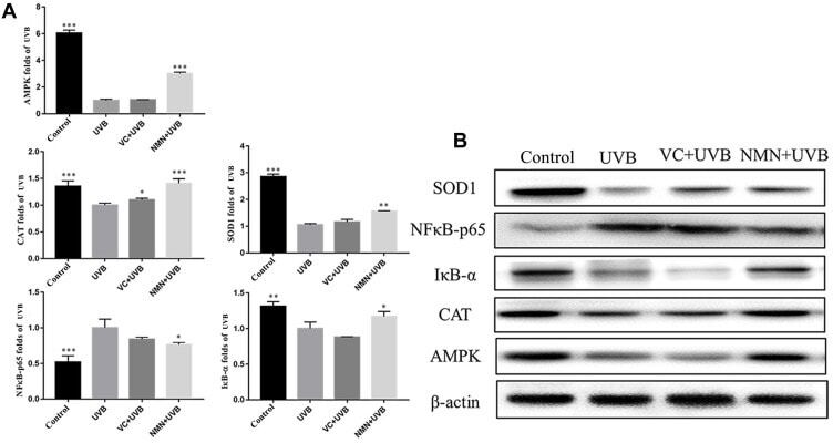
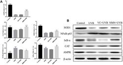
- Figure 6 Protein expression of AMPK, NFkappaB-p65, IkappaB-alpha, SOD1 and CAT in skin tissues. ( A ) relative expression levels of proteins; ( B ) protein banding map. * p < 0.05 compared to the UVB group; ** p < 0.01 compared to the UVB group; *** p < 0.001 compared to the UVB group. Abbreviations : VC+UVB, mice treated with vitamin C(300mg/kg) and UVB irradiation; NMN+UVB, mice treated with nicotinamide mononucleotide (300mg/kg) and UVB irradiation.