Explore
 Validate
 Learn
Learn Western blot
 Immunohistochemistry
Antibody data
- Antibody Data
- Antigen structure
- References [5]
- Comments [0]
- Validations
- Immunohistochemistry [3]
- Other assay [4]
Submit
Validation data
Reference
Comment
Report error
- Product number
- PA5-21078 - Provider product page

- Provider
- Invitrogen Antibodies
- Product name
- Klotho Polyclonal Antibody
- Antibody type
- Polyclonal
- Antigen
- Synthetic peptide
- Description
- A suggested positive control is HepG2 cell lysate. PA5-21078 can be used with blocking peptide PEP-1192.
- Reactivity
- Human, Mouse, Rat
- Host
- Rabbit
- Isotype
- IgG
- Vial size
- 100 μg
- Concentration
- 1 mg/mL
- Storage
- Maintain refrigerated at 2-8°C for up to 3 months. For long term storage store at -20°C
Submitted references FGF23 ameliorates ischemia-reperfusion induced acute kidney injury via modulation of endothelial progenitor cells: targeting SDF-1/CXCR4 signaling.
Towards Age-Related Anti-Inflammatory Therapy: Klotho Suppresses Activation of ER and Golgi Stress Response in Senescent Monocytes.
microRNA-200c regulates KLOTHO expression in human kidney cells under oxidative stress.
The Role of the Anti-Aging Protein Klotho in IGF-1 Signaling and Reticular Calcium Leak: Impact on the Chemosensitivity of Dedifferentiated Liposarcomas.
Protective role of klotho protein on epithelial cells upon co-culture with activated or senescent monocytes.
Chang HM, Peng KY, Chan CK, Sun CY, Chen YY, Chang HM, Huang CL, Liu PC, Chen PY, Wang KC, Wang WJ, Wu CC, Lin YF, Lai TS, Huang TM, Young GH, Lin SL, Ostermann M, Chu TS, Chueh JS, Wu VC
Cell death & disease 2021 Apr 17;12(5):409
Cell death & disease 2021 Apr 17;12(5):409
Towards Age-Related Anti-Inflammatory Therapy: Klotho Suppresses Activation of ER and Golgi Stress Response in Senescent Monocytes.
Mytych J, Sołek P, Będzińska A, Rusinek K, Warzybok A, Tabęcka-Łonczyńska A, Koziorowski M
Cells 2020 Jan 21;9(2)
Cells 2020 Jan 21;9(2)
microRNA-200c regulates KLOTHO expression in human kidney cells under oxidative stress.
Morii K, Yamasaki S, Doi S, Irifuku T, Sasaki K, Doi T, Nakashima A, Arihiro K, Masaki T
PloS one 2019;14(6):e0218468
PloS one 2019;14(6):e0218468
The Role of the Anti-Aging Protein Klotho in IGF-1 Signaling and Reticular Calcium Leak: Impact on the Chemosensitivity of Dedifferentiated Liposarcomas.
Delcroix V, Mauduit O, Tessier N, Montillaud A, Lesluyes T, Ducret T, Chibon F, Van Coppenolle F, Ducreux S, Vacher P
Cancers 2018 Nov 14;10(11)
Cancers 2018 Nov 14;10(11)
Protective role of klotho protein on epithelial cells upon co-culture with activated or senescent monocytes.
Mytych J, Wos I, Solek P, Koziorowski M
Experimental cell research 2017 Jan 15;350(2):358-367
Experimental cell research 2017 Jan 15;350(2):358-367
No comments: Submit comment
Supportive validation
- Submitted by
- Invitrogen Antibodies (provider)
- Main image
- Experimental details
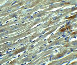
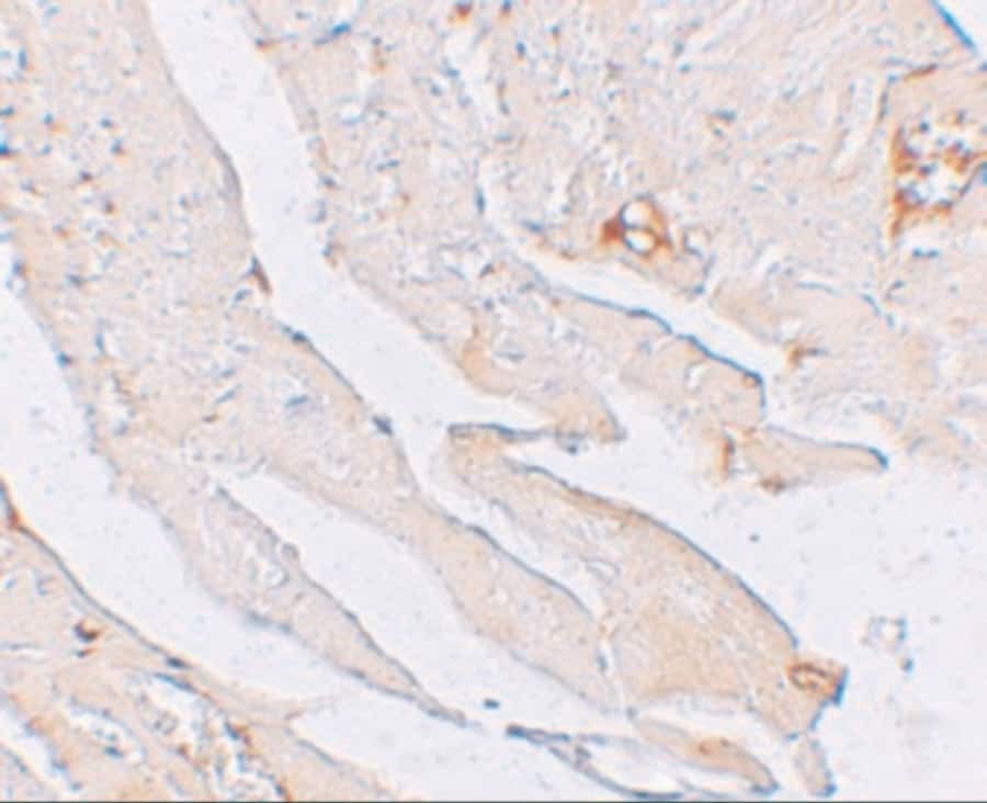
- Immunohistochemical analysis of paraffin-embedded rat heart tissue using Klotho Polyclonal Antibody (Product # PA5-21078) at 2.5 µg/mL. Tissue was fixed with formaldehyde and blocked with 0.1 serum for 1 h at RT; antigen retrieval was by heat mediation with a citrate buffer (pH6). Samples were incubated with primary antibody overnight at 4˚C. A goat anti-rabbit IgG H&L (HRP) at 1/250 was used as secondary. Counter stained with Hematoxylin.
- Submitted by
- Invitrogen Antibodies (provider)
- Main image
- Experimental details
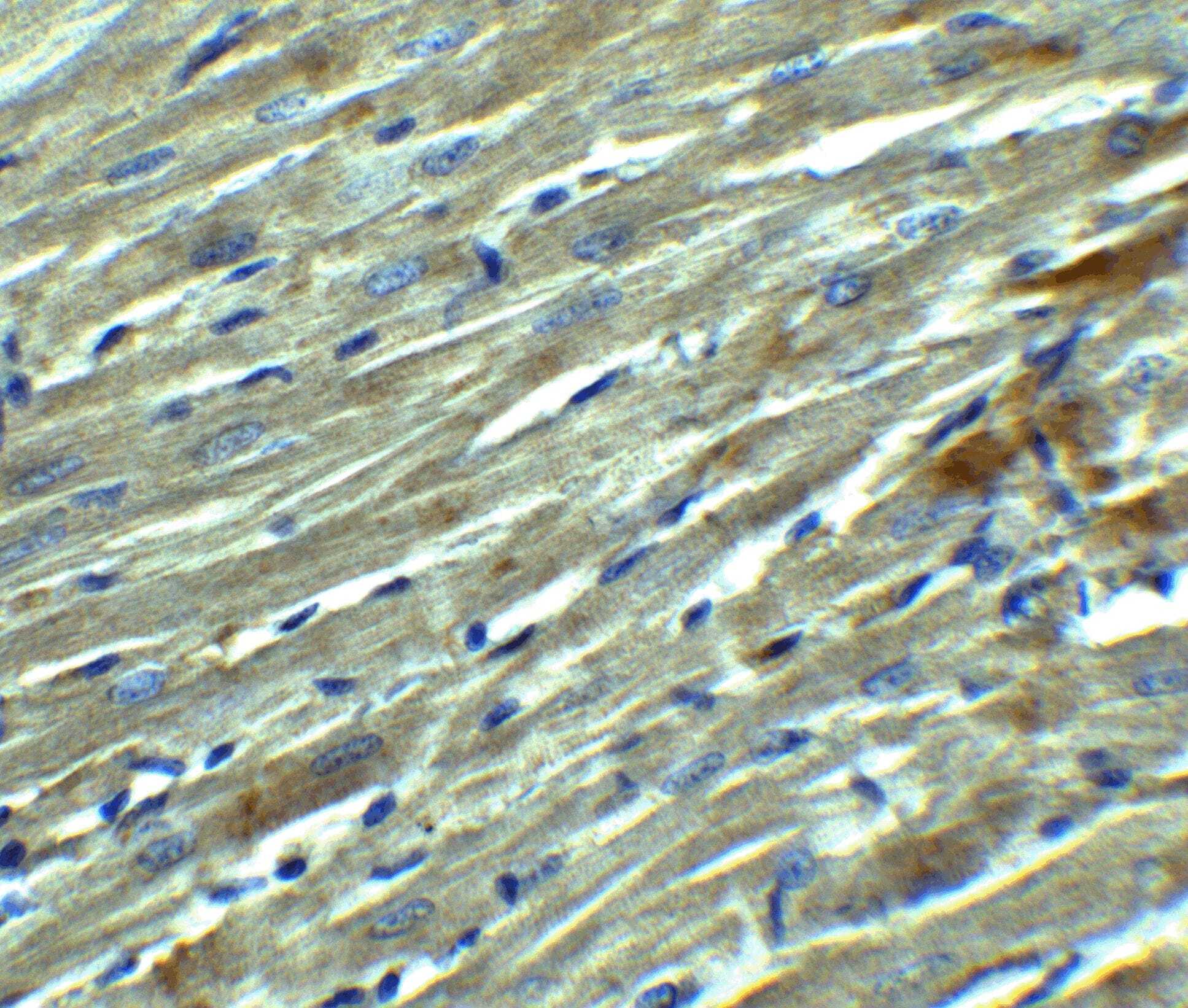
- Immunohistochemical analysis of paraffin-embedded mouse heart tissue using Klotho Polyclonal Antibody (Product # PA5-21078) at 2.5 µg/mL. Tissue was fixed with formaldehyde and blocked with 0.1 serum for 1 h at RT; antigen retrieval was by heat mediation with a citrate buffer (pH6). Samples were incubated with primary antibody overnight at 4˚C. A goat anti-rabbit IgG H&L (HRP) at 1/250 was used as secondary. Counter stained with Hematoxylin.
- Submitted by
- Invitrogen Antibodies (provider)
- Main image
- Experimental details
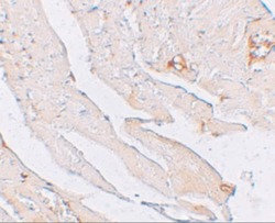
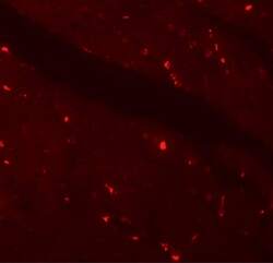
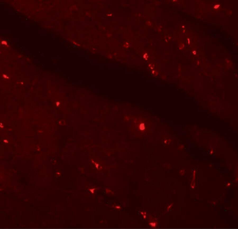
- Immunofluorescent analysis of 4% paraformaldehyde-fixed mouse heart tissue labeling KLOTHO with Klotho Polyclonal Antibody (Product # PA5-21078) at 20 µg/mL, followed by goat anti-rabbit IgG secondary antibody at 1:500 dilution (red).
Supportive validation
- Submitted by
- Invitrogen Antibodies (provider)
- Main image

- Experimental details
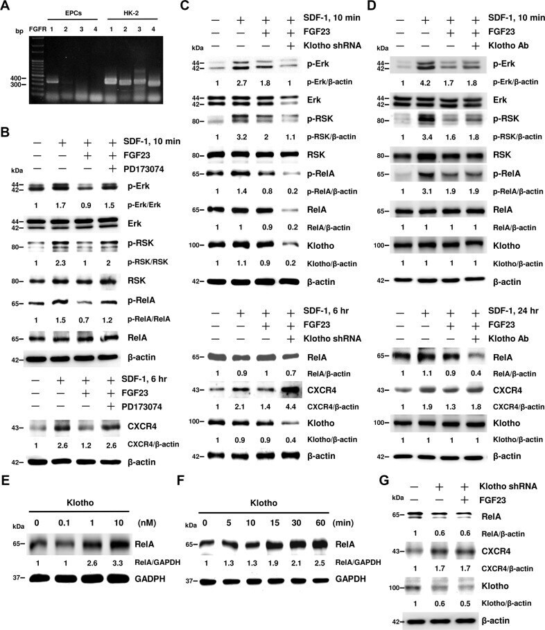
- Fig. 5 FGF23 attenuates SDF-1-induced CXCR4 expression via interaction of FGF receptor-1 (FGFR1), but not Klotho. A FGFR-1, -2, -3, or -4 mRNA expression levels in EPCs were detected by RT-PCR. HK-2 lysates were used as positive controls. B EPCs were pre-treated with FGF23 and/or FGFR inhibitor (PD173074) for 30 min followed by SDF-1 stimulation for 15 min or 6 h. C EPCs harboring control or Klotho shRNA were pre-treated with FGF23 for 30 min followed by SDF-1 stimulation for 10 min or 6 h. D EPCs were pre-treated with Klotho neutralizing antibody for 60 min and FGF23 for 30 min followed by SDF-1 stimulation for 10 min or 24 h. E EPCs were treated with different doses of Klotho. F EPCs were collected at the indicated time points after Klotho treatment. G EPCs harboring control or Klotho shRNA were pre-treated with FGF23 for 24 h. Protein lysates were analyzed by immunoblot using indicated antibodies. beta-actin or GAPDH were used as protein loading controls. The numbers under the gel lanes represent the relative protein level.
- Submitted by
- Invitrogen Antibodies (provider)
- Main image
- Experimental details
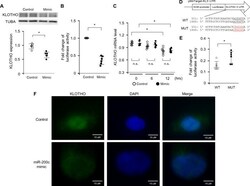
- Fig 3 miR-200c decreases KLOTHO expression in HK-2 cells by translational repression. (A) KLOTHO protein expression in HK-2 cells 24 hrs after transfection with 25 nM mimic control or miR-200c mimic. Cells were cultured for another 48 hrs without mimic control or miR-200c mimic before sampling. Band intensities were analyzed and normalized against TUBA using densitometry. (B) A KLOTHO 3'-UTR reporter plasmid in combination with 50 nM mimic control or miR-200c mimic was transfected into HK-2 cells for 4 hrs. After a medium change, HK-2 cells were cultured for another 12 hrs before sampling. Luciferase activity was normalized against protein amount. (C) HK-2 cells were transfected with 50 nM mimic control or miR-200c mimic for 4 hrs and cultured for another 24 hrs before harvesting total RNA. KLOTHO mRNA levels were evaluated by q-PCR. (D) Mutations were introduced into the 3'-UTR of KLOHO mRNA as indicated. (E) The effect of 100 nM miR-200c mimic on the reporter activity of wild type (WT, pMirTarget-KL3'-UTR-WT) and mutant (MUT, pMirTarget-KL3'-UTR-MUT) plasmids in HK-2 cells was measured by luciferase assay. (F) HK-2 cells were stained with anti-KLOTHO antibody and Alexa Fluor 488-labeled goat anti-rabbit IgG. KLOTHO protein was evaluated under fluorescence microscopy. Scale bar = 10 mum. Values represent individual measurements and the mean +- SD. Data were analyzed using the Mann-Whitney U -test or the Mann-Whitney U -test with Bonferroni correction. * P < 0.05, n

- Submitted by
- Invitrogen Antibodies (provider)
- Main image

- Experimental details
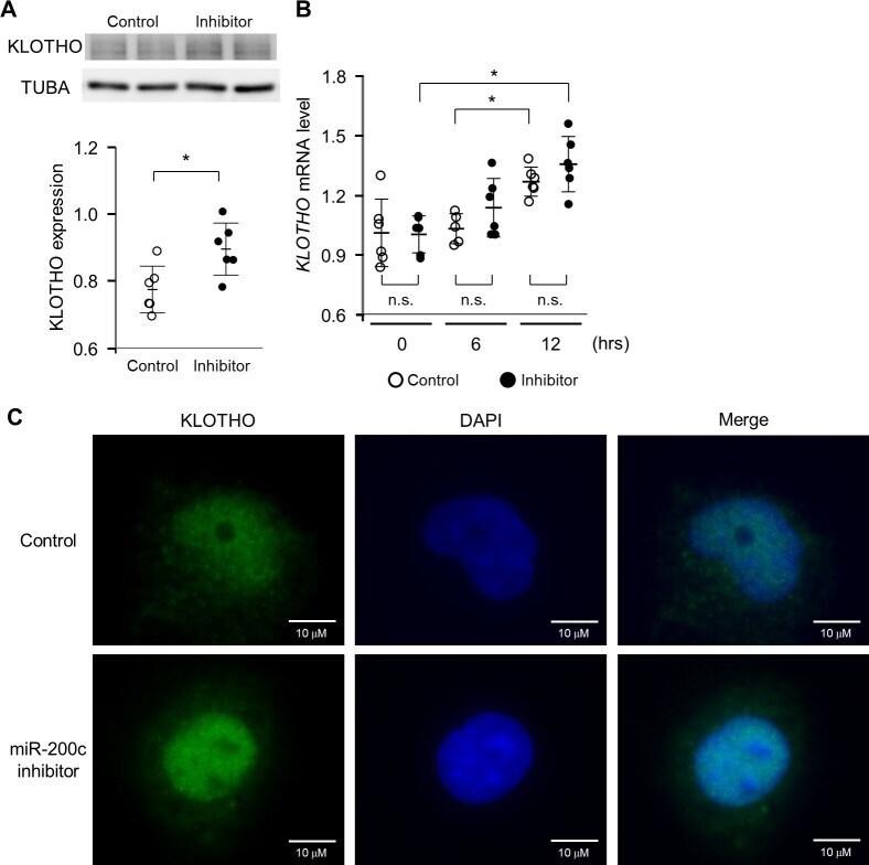
- Fig 4 KLOTHO protein is preserved by inhibiting miR-200c in HK-2 cells. (A) The effect of a miR-200c inhibitor on KLOTHO protein expression in HK-2 cells treated with H 2 O 2 . KLOTHO protein expression in HK-2 cells treated with 100 muM H 2 O 2 for 24 hrs after the transfection of inhibitor control (25 nM) or miR-200c inhibitor (25 nM) for 4 hrs. Band intensities were analyzed and normalized against TUBA using densitometry. * P < 0.05, n = 6 (B) The effect of a miR-200c inhibitor on KLOTHO mRNA expression in HK-2 cells treated with H 2 O 2 . HK-2 cells were transfected with inhibitor control (25 nM) or miR-200c inhibitor (25 nM) and 12 hrs later were treated with 100 muM H 2 O 2 . KLOTHO mRNA was detected by q-PCR. (C) HK-2 cells were stained with anti-KLOTHO antibody and Alexa Fluor 488-labeled goat anti-rabbit IgG. KLOTHO protein was evaluated under fluorescence microscopy. Scale bar = 10 mum. Values represent individual measurements and the mean +- SD. Data were analyzed using the Mann-Whitney U -test or the Mann-Whitney U -test with Bonferroni correction. * P < 0.05, n = 6. n.s.; not significant.
- Submitted by
- Invitrogen Antibodies (provider)
- Main image
- Experimental details
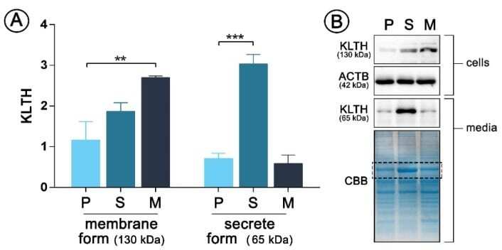
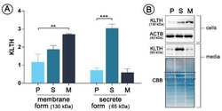
- Figure 1 pDNA(plasmid DNA)-mediated klotho overexpression in human monocytic cells Cells were transfected with pDNA and selected in antibiotics to obtain stable clones and then Western Blot analysis of klotho membrane (130 kDa) and secrete (65 kDa) forms expression was performed ( A ). Representative images of Western Blot membrane and Coomassie staining are presented ( B ). The bands were quantified and normalized to their corresponding beta-actin bands in the case of membrane form of klotho or to CBB staining in the case of secrete form. Bars indicate SD, n = 3, *** p < 0.001, ** p < 0.01 (one-way ANOVA and Dunnett's a posteriori test). ACTB actin, KLTH klotho, CBB Coomassie Brilliant Blue staining.