Explore
 Validate
 Learn
Learn Western blot
Antibody data
- Antibody Data
- Antigen structure
- References [0]
- Comments [0]
- Validations
- Western blot [1]
- ELISA [1]
- Immunohistochemistry [1]
Submit
Validation data
Reference
Comment
Report error
- Product number
- 17190002-0.1mg - Provider product page

- Provider
- Novus Biologicals
- Product name
- Rabbit Polyclonal TSP50 Antibody
- Antibody type
- Polyclonal
- Description
- Immunogen affinity purified. This product is specific for Human TSP50.
- Reactivity
- Human, Mouse
- Host
- Rabbit
- Isotype
- IgG
- Vial size
- 0.1 mg
- Storage
- Store at 4C short term. Aliquot and store at -20C long term. Avoid freeze-thaw cycles.
No comments: Submit comment
Supportive validation
- Submitted by
- Novus Biologicals (provider)
- Main image
- Experimental details
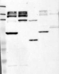
- Western Blot: TSP50 Antibody [17190002] - Samples: Lane 1, Marker [kDa]: 219, 111, 83, 48, 32, 26, 17 Lane 2, RT-4 Lane 3, U-251MG sp Lane 4, Human Plasma Lane 5, Liver Lane 6, Tonsil, Target weight [kDa]: 43 Validation score: 2 Validation description: Supportive - Band of predicted size in kDa (+/-20%) with additional bands present.

Supportive validation
- Submitted by
- Novus Biologicals (provider)
- Main image
- Experimental details
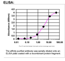
- ELISA: TSP50 Antibody [17190002]

Supportive validation
- Submitted by
- Novus Biologicals (provider)
- Main image

- Experimental details
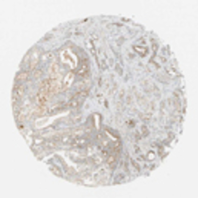
- Immunohistochemistry: TSP50 Antibody [17190002] - Strong cytoplasmic positivity was seen in primary spermatocytes of the seminiferous tubules of the testes, neurons of the cerebral cortex and many hematopoeitic cells of the bone marrow. Most mucosal surfaces and glands showed moderate or some times weak positivity. Biliary ductal cells and gall bladder mucosal cells showed strong accentuation of the luminal cell membrane. Most other normal tissues showed moderate or weak positivity. More consistent and stronger expression was seen in malignancies. Strong or moderate positivity was seen in prostatic, testicular and colo-rectal cancers. Many pancreatic, prostatic and some testicular cancers showed strong cytoplasmic granularity. Strong or moderate positivity was also seen in malignant carcinoid, renal and urothelial cancers. Most other cancers showed moderate or sometimes weak positivity. Image and statement courtesy of the Human Protein Atlas (HPA).