Explore
 Validate
 Learn
Learn Western blot
Antibody data
- Antibody Data
- Antigen structure
- References [0]
- Comments [0]
- Validations
- Western blot [5]
- Immunohistochemistry [1]
Submit
Validation data
Reference
Comment
Report error
- Product number
- PA5-70004 - Provider product page

- Provider
- Invitrogen Antibodies
- Product name
- GSR Polyclonal Antibody
- Antibody type
- Polyclonal
- Antigen
- Synthetic peptide
- Description
- This target displays homology in the following species: Cow: 93%; Dog: 93%; Guinea Pig: 100%; Horse: 86%; Human: 100%; Mouse: 100%; Rabbit: 100%; Rat: 93%
- Reactivity
- Human, Mouse, Zebrafish
- Host
- Rabbit
- Isotype
- IgG
- Vial size
- 100 µL
- Concentration
- 0.5 mg/mL
- Storage
- -20° C, Avoid Freeze/Thaw Cycles
No comments: Submit comment
Supportive validation
- Submitted by
- Invitrogen Antibodies (provider)
- Main image
- Experimental details
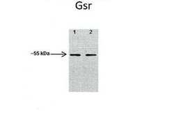
- Western blot analysis of GSR on mouse heart, mouse skeletal muscle tissue lysate using 40 µg of sample per lane. Lane 1: mouse heart, Lane 2: mouse skeletal muscle. The sample was probed with a GSR polyclonal antibody (Product # PA5-70004) using a primary antibody dilution of 1:1000 and a secondary antibody dilution of 1:10000.

- Submitted by
- Invitrogen Antibodies (provider)
- Main image
- Experimental details
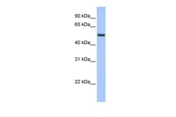
- Western blot analysis of GSR on human HeLa cells. The sample was probed with a GSR polyclonal antibody (Product # PA5-70004) using a primary antibody dilution of 0.2-1.0 µg/mL.

- Submitted by
- Invitrogen Antibodies (provider)
- Main image
- Experimental details
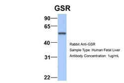
- Western blot analysis of GSR on human fetal liver tissue lysate. The sample was probed with a GSR polyclonal antibody (Product # PA5-70004) using a primary antibody dilution of 1.0 µg/mL.

- Submitted by
- Invitrogen Antibodies (provider)
- Main image
- Experimental details
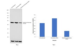
- Knockdown of GSR was achieved by transfecting Hep G2 with GSR specific siRNAs (Silencer® select Product # s6248). Western blot analysis (Fig. a) was performed using whole cell extracts from the GSR knockdown cells (lane 3), non-specific scrambled siRNA transfected cells (lane 2) and untransfected cells (lane 1). The blot was probed with GSR Polyclonal Antibody (Product # PA5-70004, 1:1000 dilution) and Goat anti-Rabbit IgG (H+L) Superclonal™ Recombinant Secondary Antibody, HRP (Product # A27036, 1:4000 dilution). Densitometric analysis of this western blot is shown in histogram (Fig. b). Decrease in signal upon siRNA mediated knock down confirms that antibody is specific to GSR.

- Submitted by
- Invitrogen Antibodies (provider)
- Main image
- Experimental details
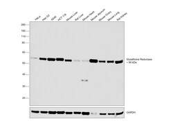
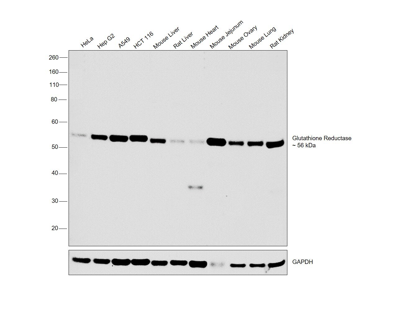
- Western blot was performed using Anti-GSR Polyclonal Antibody (Product # PA5-70004) and a band at 56 kDa corresponding to GSR was observed across the cell lines and tissues tested. Whole cell extracts (30 µg lysate) of HeLa (Lane 1), Hep G2 (Lane 2), A549 (Lane 3), HCT 116 (Lane 4), Mouse Liver (Lane 5), Rat Liver (Lane 6), Mouse Heart (Lane 7), Mouse Jejunum (Lane 8), Mouse Ovary (Lane 9), Mouse Lung (Lane 10) and Rat Kidney (Lane 11) were electrophoresed using Novex® NuPAGE® 4-12% % Bis-Tris gel (Product # NP0321BOX). Resolved proteins were then transferred onto a nitrocellulose membrane (Product # IB23001) by iBlot® 2 Dry Blotting System (Product # IB21001). The blot was probed with the primary antibody (1:1000 dilution) and detected by chemiluminescence with Goat anti-Rabbit IgG (H+L) Superclonal™ Recombinant Secondary Antibody, HRP (Product # A27036, 1:4000 dilution) using the iBright FL 1000 (Product # A32752). Chemiluminescent detection was performed using Novex® ECL Chemiluminescent Substrate Reagent Kit (Product # WP20005).
Supportive validation
- Submitted by
- Invitrogen Antibodies (provider)
- Main image

- Experimental details
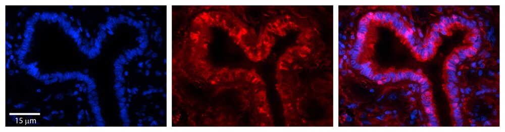
- Immunohistochemical analysis of GSR in paraffin-embedded human bronchial epithelial tissue. Sample was probed with a GSR polyclonal antibody (Product # PA5-70004) at a dilution of 1:100. Detection was performed with a donkey anti-Rabbit Cy3 secondary antibody at a dilution of 1:200. Images were taken at 20x magnification with an exposure time of 0.5-2.0 seconds.