 Explore
 Validate
 Learn
Learn Western blot
 Immunoelectron microscopy
Antibody data
- Antibody Data
- Antigen structure
- References [1]
- Comments [0]
- Validations
- Western blot [6]
- Immunohistochemistry [3]
- Other assay [1]
Submit
Validation data
Reference
Comment
Report error
- Product number
- PA5-29945 - Provider product page

- Provider
- Invitrogen Antibodies
- Product name
- GSR Polyclonal Antibody
- Antibody type
- Polyclonal
- Antigen
- Recombinant full-length protein
- Description
- Recommended positive controls: HCT116, mouse eye, mouse brain, PC-12, Rat-2. Predicted reactivity: Mouse (85%), Rat (87%), Bovine (84%). Store product as a concentrated solution. Centrifuge briefly prior to opening the vial.
- Reactivity
- Human, Mouse, Rat
- Host
- Rabbit
- Isotype
- IgG
- Vial size
- 100 μL
- Concentration
- 1.21 mg/mL
- Storage
- Store at 4°C short term. For long term storage, store at -20°C, avoiding freeze/thaw cycles.
Submitted references MYC-driven inhibition of the glutamate-cysteine ligase promotes glutathione depletion in liver cancer.
Anderton B, Camarda R, Balakrishnan S, Balakrishnan A, Kohnz RA, Lim L, Evason KJ, Momcilovic O, Kruttwig K, Huang Q, Xu G, Nomura DK, Goga A
EMBO reports 2017 Apr;18(4):569-585
EMBO reports 2017 Apr;18(4):569-585
No comments: Submit comment
Supportive validation
- Submitted by
- Invitrogen Antibodies (provider)
- Main image
- Experimental details
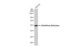
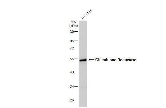
- Western Blot using GSR Polyclonal Antibody (Product # PA5-29945). Whole cell extract (30 µg) was separated by 10% SDS-PAGE, and the membrane was blotted with GSR Polyclonal Antibody (Product # PA5-29945) diluted at 1:1,000. The HRP-conjugated anti-rabbit IgG antibody was used to detect the primary antibody.
- Submitted by
- Invitrogen Antibodies (provider)
- Main image
- Experimental details
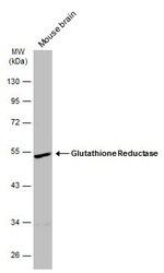
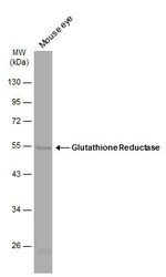
- Western blot analysis of GSR was performed by separating 50 µg of mouse tissue extract by 10% SDS-PAGE. Proteins were transferred to a membrane and probed with a GSR Polyclonal Antibody (Product # PA5-29945) at a dilution of 1:1000. The HRP-conjugated anti-rabbit IgG antibody was used to detect the primary antibody.
- Submitted by
- Invitrogen Antibodies (provider)
- Main image
- Experimental details
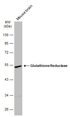
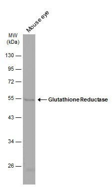
- Western blot analysis of GSR was performed by separating 50 µg of mouse tissue extract by 10% SDS-PAGE. Proteins were transferred to a membrane and probed with a GSR Polyclonal Antibody (Product # PA5-29945) at a dilution of 1:1000. The HRP-conjugated anti-rabbit IgG antibody was used to detect the primary antibody.
- Submitted by
- Invitrogen Antibodies (provider)
- Main image
- Experimental details
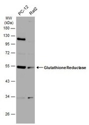
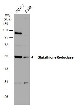
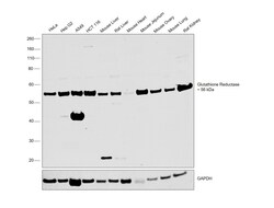
- Western Blot analysis of GSR was performed by separating 30 µg of various whole cell extracts by 10% SDS-PAGE. Proteins were transferred to a membrane and probed with a GSR Polyclonal Antibody (Product # PA5-29945) at a dilution of 1:1000 and a HRP-conjugated anti-rabbit IgG secondary antibody.
- Submitted by
- Invitrogen Antibodies (provider)
- Main image
- Experimental details
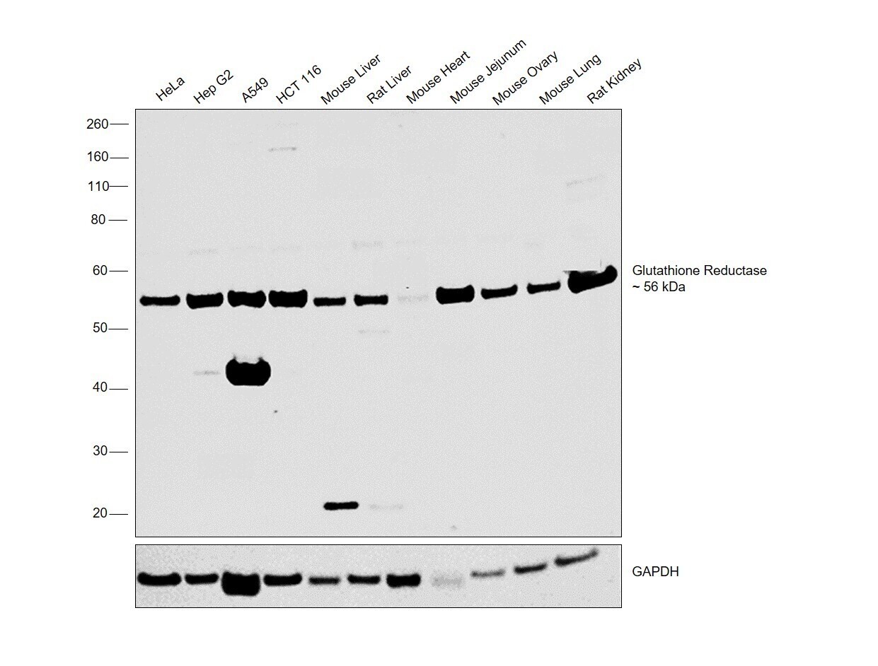
- Western blot was performed using Anti-GSR Polyclonal Antibody (Product # PA5-29945) and a band at 56 kDa corresponding to GSR was observed across the cell lines and tissues tested. Whole cell extracts (30 µg lysate) of HeLa (Lane 1), Hep G2 (Lane 2), A549 (Lane 3), HCT 116 (Lane 4), Mouse Liver (Lane 5), Rat Liver (Lane 6), Mouse Heart (Lane 7), Mouse Jejunum (Lane 8), Mouse Ovary (Lane 9), Mouse Lung (Lane 10) and Rat Kidney (Lane 11) were electrophoresed using Novex® NuPAGE® 4-12% % Bis-Tris gel (Product # NP0321BOX). Resolved proteins were then transferred onto a nitrocellulose membrane (Product # IB23001) by iBlot® 2 Dry Blotting System (Product # IB21001). The blot was probed with the primary antibody (1:1000 dilution) and detected by chemiluminescence with Goat anti-Rabbit IgG (Heavy Chain) Superclonal™ Recombinant Secondary Antibody, HRP (Product # A27036, 1:4000 dilution) using the iBright FL 1000 (Product # A32752). Chemiluminescent detection was performed using Novex® ECL Chemiluminescent Substrate Reagent Kit (Product # WP20005).
- Submitted by
- Invitrogen Antibodies (provider)
- Main image
- Experimental details
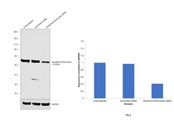
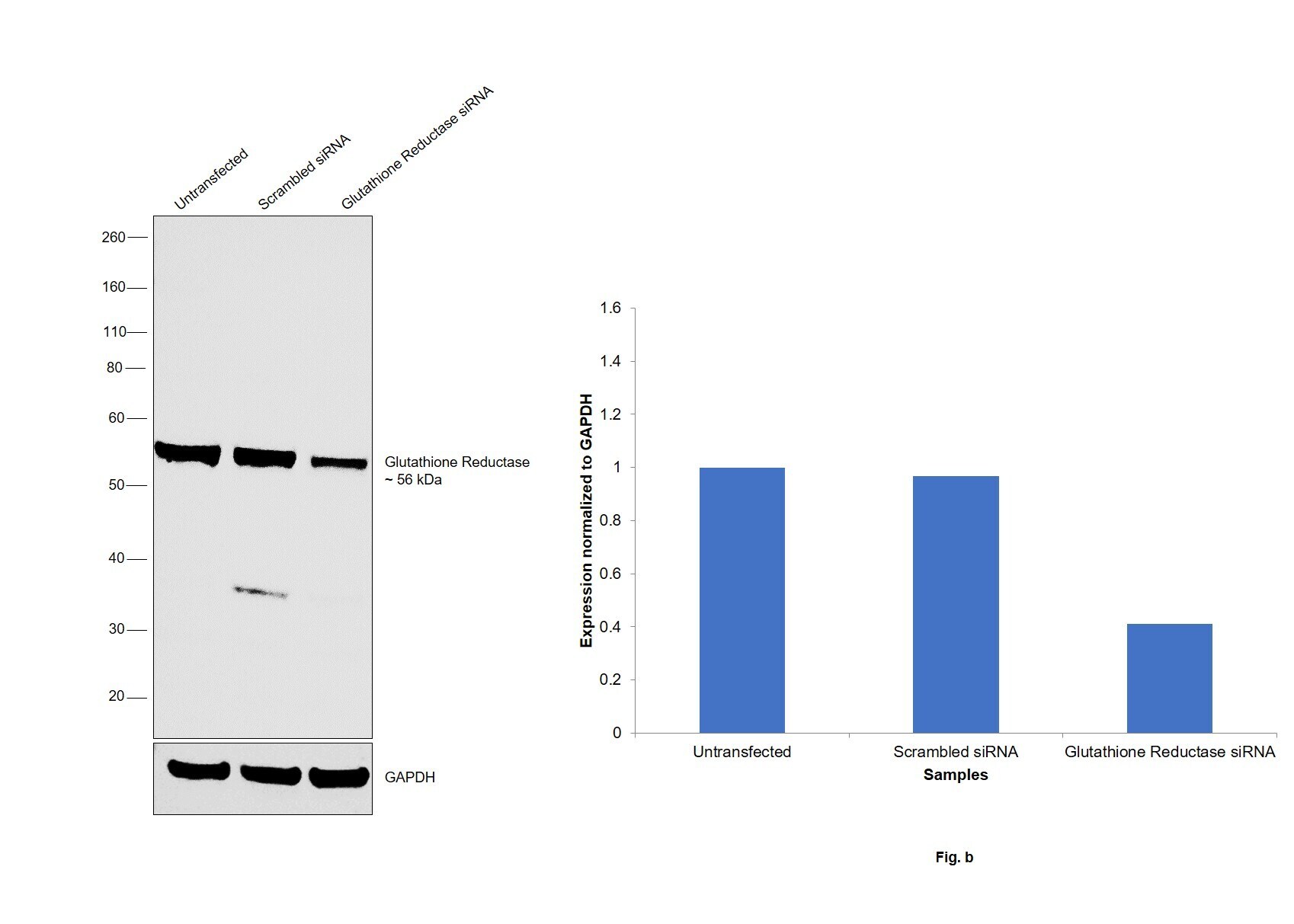
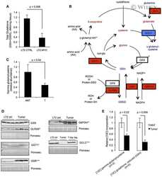
- Knockdown of GSR was achieved by transfecting Hep G2 with GSR specific siRNAs (Silencer® select Product # s6248). Western blot analysis (Fig. a) was performed using whole cell extracts from the GSR knockdown cells (lane 3), non-specific scrambled siRNA transfected cells (lane 2) and untransfected cells (lane 1). The blot was probed with GSR Polyclonal Antibody (Product # PA5-29945, 1:1000 dilution) and Goat anti-Rabbit IgG (Heavy Chain) Superclonal™ Recombinant Secondary Antibody, HRP (Product # A27036, 1:4000 dilution). Densitometric analysis of this western blot is shown in histogram (Fig. b). Decrease in signal upon siRNA mediated knock down confirms that antibody is specific to GSR.
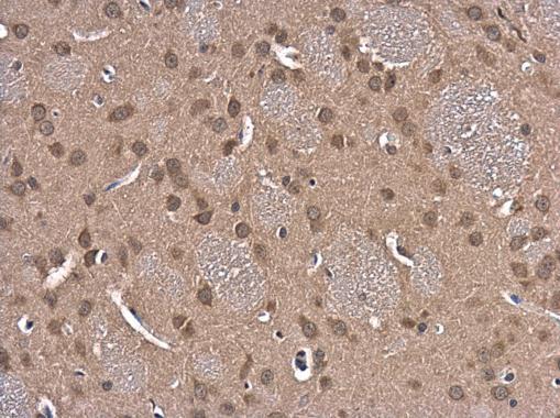
Supportive validation
- Submitted by
- Invitrogen Antibodies (provider)
- Main image
- Experimental details
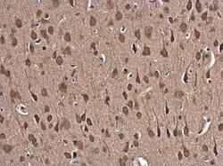
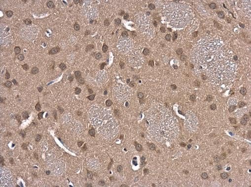
- Immunohistochemistry (Paraffin) analysis of GSR was performed in paraffin-embedded rat brain tissue using GSR Polyclonal Antibody (Product # PA5-29945) at a dilution of 1:500.

- Submitted by
- Invitrogen Antibodies (provider)
- Main image
- Experimental details
- Immunohistochemistry (Paraffin) analysis of GSR was performed in paraffin-embedded rat brain tissue using GSR Polyclonal Antibody (Product # PA5-29945) at a dilution of 1:500.
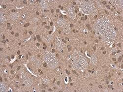
- Submitted by
- Invitrogen Antibodies (provider)
- Main image
- Experimental details
- Immunohistochemistry (Paraffin) analysis of GSR was performed in paraffin-embedded rat brain tissue using GSR Polyclonal Antibody (Product # PA5-29945) at a dilution of 1:500.
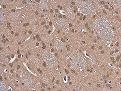
Supportive validation
- Submitted by
- Invitrogen Antibodies (provider)
- Main image
- Experimental details
- NULL
