Explore
 Validate
 Learn
Learn Western blot
 Immunocytochemistry
Antibody data
- Antibody Data
- Antigen structure
- References [2]
- Comments [0]
- Validations
- Immunocytochemistry [1]
Submit
Validation data
Reference
Comment
Report error
- Product number
- HPA005633 - Provider product page

- Provider
- Atlas Antibodies
- Proper citation
- Atlas Antibodies Cat#HPA005633, RRID:AB_1078954
- Product name
- Anti-GATA2
- Antibody type
- Polyclonal
- Description
- Polyclonal Antibody against Human GATA2, Gene description: GATA binding protein 2, Alternative Gene Names: NFE1B, Validated applications: IHC, ICC, WB, Uniprot ID: P23769, Storage: Store at +4°C for short term storage. Long time storage is recommended at -20°C.
- Reactivity
- Human
- Host
- Rabbit
- Conjugate
- Unconjugated
- Isotype
- IgG
- Vial size
- 100 µl
- Concentration
- 0.2 mg/ml
- Storage
- Store at +4°C for short term storage. Long time storage is recommended at -20°C.
- Handling
- The antibody solution should be gently mixed before use.
Submitted references GATA2 and Progesterone Receptor Interaction in Endometrial Stromal Cells Undergoing Decidualization
Nuclear Pores Promote Lethal Prostate Cancer by Increasing POM121-Driven E2F1, MYC, and AR Nuclear Import
Kohlmeier A, Sison C, Yilmaz B, Coon V J, Dyson M, Bulun S
Endocrinology 2020;161(6)
Endocrinology 2020;161(6)
Nuclear Pores Promote Lethal Prostate Cancer by Increasing POM121-Driven E2F1, MYC, and AR Nuclear Import
Rodriguez-Bravo V, Pippa R, Song W, Carceles-Cordon M, Dominguez-Andres A, Fujiwara N, Woo J, Koh A, Ertel A, Lokareddy R, Cuesta-Dominguez A, Kim R, Rodriguez-Fernandez I, Li P, Gordon R, Hirschfield H, Prats J, Reddy E, Fatatis A, Petrylak D, Gomella L, Kelly W, Lowe S, Knudsen K, Galsky M, Cingolani G, Lujambio A, Hoshida Y, Domingo-Domenech J
Cell 2018;174(5):1200-1215.e20
Cell 2018;174(5):1200-1215.e20
No comments: Submit comment
Supportive validation
- Submitted by
- Atlas Antibodies (provider)
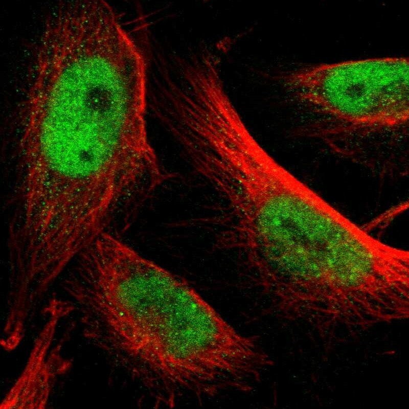
- Main image

- Experimental details
- Immunofluorescent staining of human cell line U-251 MG shows localization to nucleoplasm.
- Sample type
- Human