 Explore
 Validate
 Learn
Learn Western blot
 Immunocytochemistry
 Immunoprecipitation
Antibody data
- Antibody Data
- Antigen structure
- References [1]
- Comments [0]
- Validations
- Immunocytochemistry [7]
- Immunohistochemistry [2]
- Other assay [1]
Submit
Validation data
Reference
Comment
Report error
- Product number
- MA5-24860 - Provider product page

- Provider
- Invitrogen Antibodies
- Product name
- DGCR8 Recombinant Rabbit Monoclonal Antibody (3F5)
- Antibody type
- Monoclonal
- Antigen
- Recombinant full-length protein
- Description
- Recombinant rabbit monoclonal antibodies are produced using in vitro expression systems. The expression systems are developed by cloning in the specific antibody DNA sequences from immunoreactive rabbits. Then, individual clones are screened to select the best candidates for production. The advantages of using recombinant rabbit monoclonal antibodies include: better specificity and sensitivity, lot-to-lot consistency, animal origin-free formulations, and broader immunoreactivity to diverse targets due to larger rabbit immune repertoire. Purity is > 95% by SDS-PAGE.
- Reactivity
- Human, Mouse, Rat
- Host
- Rabbit
- Isotype
- IgG
- Antibody clone number
- 3F5
- Vial size
- 100 μL
- Concentration
- 1 mg/mL
- Storage
- Store at 4°C short term. For long term storage, store at -20°C, avoiding freeze/thaw cycles.
Submitted references Small RNA expression and miRNA modification dynamics in human oocytes and early embryos.
Paloviita P, Hydén-Granskog C, Yohannes DA, Paluoja P, Kere J, Tapanainen JS, Krjutškov K, Tuuri T, Võsa U, Vuoristo S
Genome research 2021 Aug;31(8):1474-1485
Genome research 2021 Aug;31(8):1474-1485
No comments: Submit comment
Supportive validation
- Submitted by
- Invitrogen Antibodies (provider)
- Main image
- Experimental details
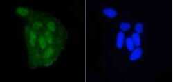
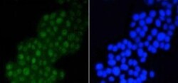
- Immunofluorescence analysis of DGCR8 in HeLa cells (green). Samples were treated with paraformaldehyde, permeabilized with 0.25% Triton and PBS, and incubated with DGCR8 monoclonal antibody (Product # MA5-24860).
- Submitted by
- Invitrogen Antibodies (provider)
- Main image

- Experimental details
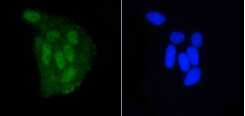
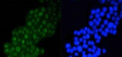
- Immunofluorescence analysis of DGCR8 in PC12 cells (green). Samples were treated with paraformaldehyde, permeabilized with 0.25% Triton and PBS, and incubated with DGCR8 monoclonal antibody (Product # MA5-24860).

- Submitted by
- Invitrogen Antibodies (provider)
- Main image

- Experimental details
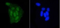
- Immunofluorescence analysis of DGCR8 in HeLa cells (green). Samples were treated with paraformaldehyde, permeabilized with 0.25% Triton and PBS, and incubated with DGCR8 monoclonal antibody (Product # MA5-24860).

- Submitted by
- Invitrogen Antibodies (provider)
- Main image
- Experimental details
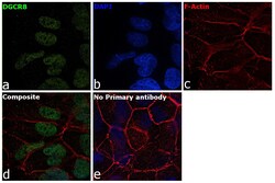
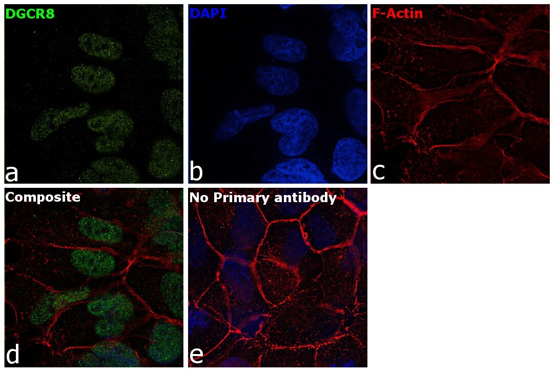
- Immunofluorescence analysis of DGCR8 was performed using HEK-293 cells. The cells were fixed with 4% paraformaldehyde for 10 minutes, permeabilized with 0.1% Triton™ X-100 for 15 minutes, and blocked with 2% BSA for 1 hour at room temperature. The cells were labeled with DGCR8 Polyclonal Antibody (Product # MA5-24860) at 1:100 dilution in 0.1% BSA and incubated overnight at 4 degree and then labeled with Donkey anti-Rabbit IgG (H+L) Highly Cross-Adsorbed Secondary Antibody, Alexa Fluor Plus 488 conjugate (Product # A32790) at a dilution of 1:2000 for 45 minutes at room temperature (Panel a: green). Nuclei (Panel b: blue) were stained with ProLong™ Diamond Antifade Mountant with DAPI (Product # P36962). F-actin (Panel c: red) was stained with Rhodamine Phalloidin (Product # R415, 1:300). Panel d represents the composite image showing nuclear localization of DGCR8 in HEK-293 cells. Panel e represents control cells with no primary antibody to assess background. The images were captured at 60X magnification.
- Submitted by
- Invitrogen Antibodies (provider)
- Main image
- Experimental details
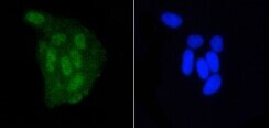
- Immunofluorescence analysis of DGCR8 in PC12 cells (green). Samples were treated with paraformaldehyde, permeabilized with 0.25% Triton and PBS, and incubated with DGCR8 monoclonal antibody (Product # MA5-24860).
- Submitted by
- Invitrogen Antibodies (provider)
- Main image
- Experimental details
- Immunofluorescence analysis of DGCR8 in HeLa cells (green). Samples were treated with paraformaldehyde, permeabilized with 0.25% Triton and PBS, and incubated with DGCR8 monoclonal antibody (Product # MA5-24860).
- Submitted by
- Invitrogen Antibodies (provider)
- Main image

- Experimental details
- Immunofluorescence analysis of DGCR8 was performed using HEK-293 cells. The cells were fixed with 4% paraformaldehyde for 10 minutes, permeabilized with 0.1% Triton™ X-100 for 15 minutes, and blocked with 2% BSA for 1 hour at room temperature. The cells were labeled with DGCR8 Polyclonal Antibody (Product # MA5-24860) at 1:100 dilution in 0.1% BSA and incubated overnight at 4 degree and then labeled with Donkey anti-Rabbit IgG (H+L) Highly Cross-Adsorbed Secondary Antibody, Alexa Fluor Plus 488 conjugate (Product # A32790) at a dilution of 1:2000 for 45 minutes at room temperature (Panel a: green). Nuclei (Panel b: blue) were stained with ProLong™ Diamond Antifade Mountant with DAPI (Product # P36962). F-actin (Panel c: red) was stained with Rhodamine Phalloidin (Product # R415, 1:300). Panel d represents the composite image showing nuclear localization of DGCR8 in HEK-293 cells. Panel e represents control cells with no primary antibody to assess background. The images were captured at 60X magnification.

Supportive validation
- Submitted by
- Invitrogen Antibodies (provider)
- Main image

- Experimental details
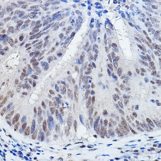
- Immunohistochemistry analysis of DGCR8 in paraffin-embedded human colon carcinoma. Samples were incubated with DGCR8 polyclonal antibody (Product # MA5-24860) using a dilution of 1:200. Perform high pressure antigen retrieval with 10 mM citrate buffer pH 6.0 before commencing with IHC staining protocol.
- Submitted by
- Invitrogen Antibodies (provider)
- Main image
- Experimental details
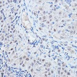
- Immunohistochemistry analysis of DGCR8 in paraffin-embedded rat ovary. Samples were incubated with DGCR8 polyclonal antibody (Product # MA5-24860) using a dilution of 1:200. Perform high pressure antigen retrieval with 10 mM citrate buffer pH 6.0 before commencing with IHC staining protocol.
Supportive validation
- Submitted by
- Invitrogen Antibodies (provider)
- Main image
- Experimental details
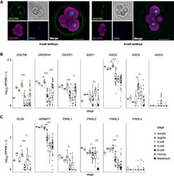
- Figure 1. Small RNA (sRNA) pathway component expression in human oocytes and embryos. ( A ) Human four-cell ( left ) and eight-cell ( right ) embryos immunostained with DGCR8 (green) and DICER1 (magenta) antibodies. Nuclei are counterstained with DAPI (blue). Overlay of a single representative z -plane and the corresponding z -planes are shown. Nucleus and bright field channels are on the right side of each overlay. Scale bar is 50 mum. Expression levels (RPKM) in human oocytes and preimplantation embryos (): ( B ) miRNA pathway gene, DGCR8 , DROSHA , DICER1 , and AGO1-4 ; ( C ) piRNA pathway gene, PLD6 , HENMT1 , and PIWIL1-4 . ( B , C ) Significant expression changes between consecutive developmental stages were assessed using pairwise, two-sided Wilcoxon rank-sum tests: (*) FDR < 0.05; (**) FDR < 0.01; (***) FDR < 0.001. The horizontal line in the box plot indicates the expression median.
