Explore
 Validate
 Learn
Learn Western blot
Antibody data
- Antibody Data
- Antigen structure
- References [7]
- Comments [0]
- Validations
- Western blot [1]
- Immunohistochemistry [3]
- Other assay [4]
Submit
Validation data
Reference
Comment
Report error
- Product number
- PA5-16634 - Provider product page

- Provider
- Invitrogen Antibodies
- Product name
- VWF Polyclonal Antibody
- Antibody type
- Polyclonal
- Antigen
- Purifed from natural sources
- Description
- PA5-16634 targets von Willebrand Factor in IF and IHC (P) applications and shows reactivity with Human samples.
Submitted references Non-invasive administration of AAV to target lung parenchymal cells and develop SARS-CoV-2-susceptible mice.
Infarct-preconditioning exosomes of umbilical cord mesenchymal stem cells promoted vascular remodeling and neurological recovery after stroke in rats.
Myeloid lineage contributes to pathological choroidal neovascularization formation via SOCS3.
Dual roles of p62/SQSTM1 in the injury and recovery phases of acetaminophen-induced liver injury in mice.
Increased talin-vinculin spatial proximities in livers in response to spotted fever group rickettsial and Ebola virus infections.
Identification of a pro-angiogenic functional role for FSP1-positive fibroblast subtype in wound healing.
Efficient One-Step Production of Microencapsulated Hepatocyte Spheroids with Enhanced Functions.
Yang MS, Park MJ, Lee J, Oh B, Kang KW, Kim Y, Lee SM, Lim JO, Jung TY, Park JH, Park SC, Lim YS, Hwang SB, Lyoo KS, Kim DI, Kim B
Molecular therapy : the journal of the American Society of Gene Therapy 2022 May 4;30(5):1994-2004
Molecular therapy : the journal of the American Society of Gene Therapy 2022 May 4;30(5):1994-2004
Infarct-preconditioning exosomes of umbilical cord mesenchymal stem cells promoted vascular remodeling and neurological recovery after stroke in rats.
Ye YC, Chang ZH, Wang P, Wang YW, Liang J, Chen C, Wang JJ, Sun HT, Wang Y, Li XH
Stem cell research & therapy 2022 Jul 28;13(1):378
Stem cell research & therapy 2022 Jul 28;13(1):378
Myeloid lineage contributes to pathological choroidal neovascularization formation via SOCS3.
Wang T, Zhou P, Xie X, Tomita Y, Cho S, Tsirukis D, Lam E, Luo HR, Sun Y
EBioMedicine 2021 Nov;73:103632
EBioMedicine 2021 Nov;73:103632
Dual roles of p62/SQSTM1 in the injury and recovery phases of acetaminophen-induced liver injury in mice.
Qian H, Bai Q, Yang X, Akakpo JY, Ji L, Yang L, Rülicke T, Zatloukal K, Jaeschke H, Ni HM, Ding WX
Acta pharmaceutica Sinica. B 2021 Dec;11(12):3791-3805
Acta pharmaceutica Sinica. B 2021 Dec;11(12):3791-3805
Increased talin-vinculin spatial proximities in livers in response to spotted fever group rickettsial and Ebola virus infections.
Liu Y, Xiao J, Zhang B, Shelite TR, Su Z, Chang Q, Judy B, Li X, Drelich A, Bei J, Zhou Y, Zheng J, Jin Y, Rossi SL, Tang SJ, Wakamiya M, Saito T, Ksiazek T, Kaphalia B, Gong B
Laboratory investigation; a journal of technical methods and pathology 2020 Aug;100(8):1030-1041
Laboratory investigation; a journal of technical methods and pathology 2020 Aug;100(8):1030-1041
Identification of a pro-angiogenic functional role for FSP1-positive fibroblast subtype in wound healing.
Saraswati S, Marrow SMW, Watch LA, Young PP
Nature communications 2019 Jul 9;10(1):3027
Nature communications 2019 Jul 9;10(1):3027
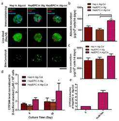
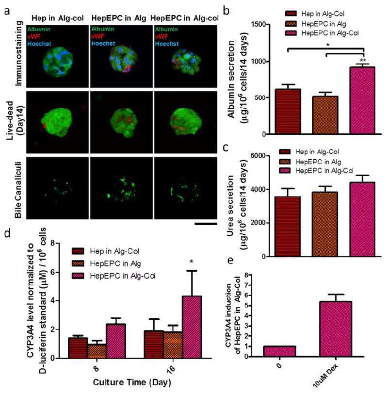
Efficient One-Step Production of Microencapsulated Hepatocyte Spheroids with Enhanced Functions.
Chan HF, Zhang Y, Leong KW
Small (Weinheim an der Bergstrasse, Germany) 2016 May;12(20):2720-30
Small (Weinheim an der Bergstrasse, Germany) 2016 May;12(20):2720-30
No comments: Submit comment
Supportive validation
- Submitted by
- Invitrogen Antibodies (provider)
- Main image
- Experimental details
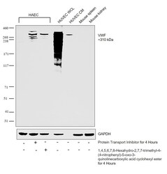
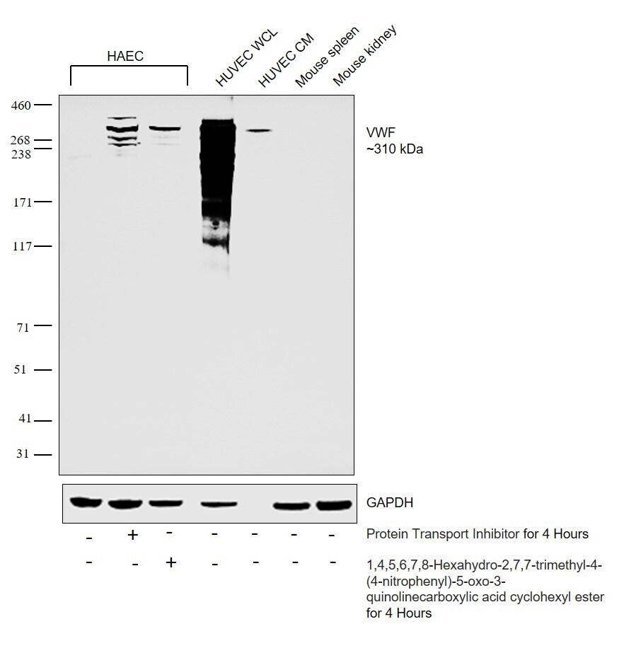
- Western blot was performed using Anti-VWF Rabbit Polyclonal Antibody (Product # PA5-16634) and a 310 kDa band corresponding to processed secretory form of VWF and cellular fragments (120-260 kDa) were observed across cell lines tested. Whole cell extracts (30 µg lysate) of HAEC (Lane 1), HAEC treated with Protein transport inhibitor (1X) (Lane 2), HAEC treated with 1,4,5,6,7,8-Hexahydro-2,7,7-trimethyl-4-(4-nitrophenyl)-5-oxo-3-quinolinecarboxylic acid cyclohexyl ester (Lane 3), HUVEC (Lane 4), HUVEC conditioned medium (Lane 5), Mouse spleen (Lane 6) and Mouse kidney (Lane 7) were electrophoresed using Novex® NuPAGE® 4-12 % Bis-Tris gel (Product # NP0322BOX). Resolved proteins were then transferred onto a nitrocellulose membrane (Product # IB23001) by iBlot® 2 Dry Blotting System (Product # IB21001). The blots were probed with the primary antibody (2 µg/mL) and detected by chemiluminescence Goat Anti-Rabbit IgG Secondary Antibody, HRP conjugate (Product # A27036, 1:4000 dilution) using the iBright FL 1000 (Product # A32752). Chemiluminescent detection was performed using Novex® ECL Chemiluminescent Substrate Reagent Kit (Product # WP20005).
Supportive validation
- Submitted by
- Invitrogen Antibodies (provider)
- Main image
- Experimental details
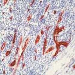
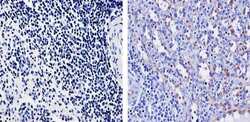
- Formalin-fixed, paraffin-embedded human tonsil stained with Factor VIII antibody using peroxidase-conjugate and AEC chromogen. Note cytoplasmic staining of endothelial cells.

- Submitted by
- Invitrogen Antibodies (provider)
- Main image
- Experimental details
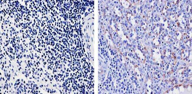
- Immunohistochemistry analysis of von Willebrand Factor showing secreted staining of paraffin-embedded human spleen tissue (right) compared to a negative control without primary antibody (left). To expose target proteins, antigen retrieval was performed using 10mM sodium citrate (pH 6.0), microwaved for 8-15 min. Following antigen retrieval, tissues were blocked in 3% H2O2-methanol for 15 min at room temperature, washed with ddH2O and PBS, and then probed with a von Willebrand Factor Rabbit Polyclonal Antibody (Product # PA5-16634) diluted in 3% BSA-PBS at a dilution of 1:100 for 1 hour at 37ºC in a humidified chamber. Tissues were washed extensively in PBST and detection was performed using an HRP-conjugated secondary antibody followed by colorimetric detection using a DAB kit. Tissues were counterstained with hematoxylin and dehydrated with ethanol and xylene to prep for mounting.
- Submitted by
- Invitrogen Antibodies (provider)
- Main image
- Experimental details
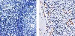
- Immunohistochemistry analysis of von Willebrand Factor showing secreted staining of paraffin-embedded human tonsil tissue (right) compared to a negative control without primary antibody (left). To expose target proteins, antigen retrieval was performed using 10mM sodium citrate (pH 6.0), microwaved for 8-15 min. Following antigen retrieval, tissues were blocked in 3% H2O2-methanol for 15 min at room temperature, washed with ddH2O and PBS, and then probed with a von Willebrand Factor Rabbit Polyclonal Antibody (Product # PA5-16634) diluted in 3% BSA-PBS at a dilution of 1:100 for 1 hour at 37ºC in a humidified chamber. Tissues were washed extensively in PBST and detection was performed using an HRP-conjugated secondary antibody followed by colorimetric detection using a DAB kit. Tissues were counterstained with hematoxylin and dehydrated with ethanol and xylene to prep for mounting.
Supportive validation
- Submitted by
- Invitrogen Antibodies (provider)
- Main image
- Experimental details
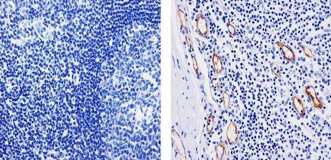
- NULL
- Submitted by
- Invitrogen Antibodies (provider)
- Main image
- Experimental details
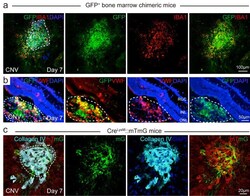
- Fig. 1 Myeloid lineage contributed to pathological endothelium formation in mice with laser-induced CNV. (a) GFP bone marrow chimeric mice were subjected to laser-induced CNV model and choroid flat mounts at Day seven after laser were stained with macrophage/microglia marker, IBA1 (red). (b) GFP bone marrow chimeric mice were subjected to laser-induced CNV and retinal cross sections at Day seven after laser were stained with EC marker vWF (red). White dotted line indicated the CNV lesion areas on retinal cross sections; (c) 8-week-old myeloid-specific LysM Cre driven mTmG reporter mice were subjected to laser-induced CNV model and choroid flat mounts at Day seven after laser were stained with EC marker Collagen IV (cyan). White dotted line indicated the CNV lesion areas on choroid flat mount. mG: membrane GFP; mT: membrane Tomato red. Scale bar: 100um in (a), 50um in (b), and 20um in (c). Fig. 1

- Submitted by
- Invitrogen Antibodies (provider)
- Main image
- Experimental details
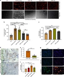
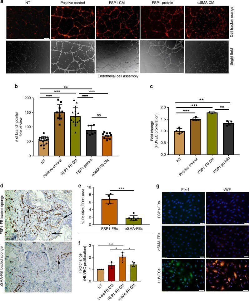
- Fig. 7 FSP1 + fibroblasts are pro-angiogenic in vitro and in vivo. a Representative images from an endothelial cell assembly assay using HUVECs treated with conditioned media (CM) collected from fibroblasts (P3-P5) or FSP1 recombinant protein. In the top row, the endothelial cells were stained with cell tracker orange and the second row contains corresponding bright field images (x20). Scale bar = 100 nM. b Quantification from the endothelial cell assembly assay based on the number of intersecting branch points per field of view (x10). Data represent an average of HUVEC cell assembly from n = 16 for NT, n = 10 for positive control, n = 15 for FSP1 + FB CM, n = 7 for FSP1 protein, and n = 10 for alphaSMA + FB CM images from three independent biological replicates; bar represents mean +- SD. c BrdU proliferation assay performed on HUVECs (P5) in response to either FSP1 + fibroblasts conditioned media (with 0.2% serum; n = 3) or FSP1 recombinant protein (10 nM; n = 4). HUVECs cultured in full serum (2%; n = 4) were used as positive control. d Representative images of FSP1 + and alphaSMA + fibroblast soak loaded sponges stained with CD31 to analyze vascular density. SP = sponge matrix, arrows point at positive stain. Scale bar = 500 uM. e Vascular density graphed as percentage of immunopositive CD31 area/total tissue area in histologic sections from granulation tissue. Data represent averages of multiple 40x fields from unpaired samples ( n = 6 fo
- Submitted by
- Invitrogen Antibodies (provider)
- Main image
- Experimental details
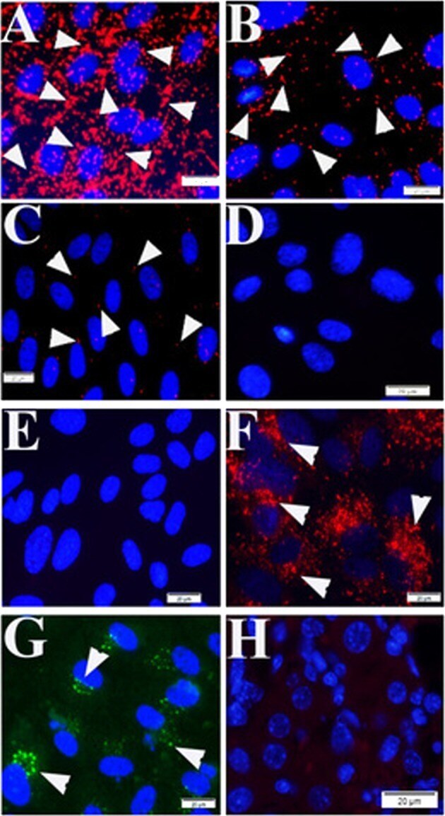
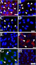
- Fig. 1 Positive and negative controls for the PLAs. Signal positive ( a-c ) and signal negative ( d ) controls of the proximity ligation assays (PLA) were established using HUVECs. Mouse anti-talin antibodies were titrated at 1:50 ( a ), 1:500 ( b ), and 1:2000 ( c ) during PLA assay. There is no positive signal visualized during PLA using mouse anti-Rab5 and rabbit anti-vWF in HUVECs ( d ) and mouse liver tissue ( h ). Normal mouse and rabbit IgGs were used as reagent negative controls during PLA assay in HUVECs ( e ). IF to von Willebrand factor (arrowheads in f ) and Rab5 (arrowheads in g ) were processed in HUVECs. Nuclei were counter-stained with DAPI (blue). Scale bars indicate 20 um.