Explore
 Validate
 Learn
Learn14-0628-80
antibody from Invitrogen Antibodies
Targeting: SELP
CD62, CD62P, GMP140, GRMP, PADGEM, PSEL
 Immunocytochemistry
Antibody data
- Antibody Data
- Antigen structure
- References [3]
- Comments [0]
- Validations
- Immunocytochemistry [1]
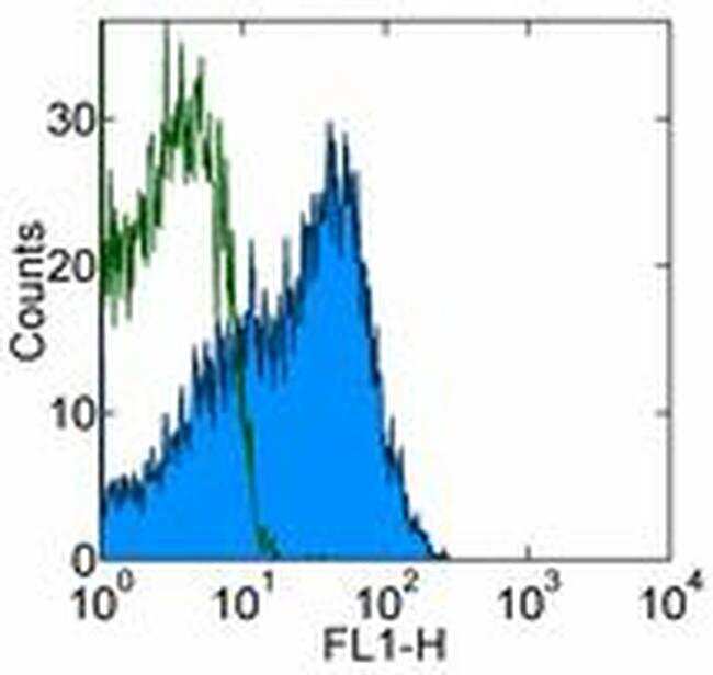
- Flow cytometry [1]
Submit
Validation data
Reference
Comment
Report error
- Product number
- 14-0628-80 - Provider product page

- Provider
- Invitrogen Antibodies
- Product name
- CD62P (P-Selectin) Monoclonal Antibody (AK-4), eBioscience™
- Antibody type
- Monoclonal
- Antigen
- Other
- Description
- Description: The AK-4 monoclonal antibody reacts with human CD62P, a 140 kDa transmembrane glycoprotein also known as P-Selectin. Upon activation, CD62P is rapidly transported from its cytoplasmic stores in the Weibel-Palade bodies of endothelial cells or the alpha-granules of platelets to the surface. CD62P initiates the adhesive interaction between endothelial cells and neutrophils and monocytes during the inflammatory reaction and is also involved in the interaction of platelets with monocytes and neutrophils.
- Antibody clone number
- AK-4
- Concentration
- 0.5 mg/mL
Submitted references Soluble Siglec-5 associates to PSGL-1 and displays anti-inflammatory activity.
Monitoring receptor-ligand interactions between surfaces by thermal fluctuations.
GMP-140 binding to neutrophils is inhibited by sulfated glycans.
Pepin M, Mezouar S, Pegon J, Muczynski V, Adam F, Bianchini EP, Bazaa A, Proulle V, Rupin A, Paysant J, Panicot-Dubois L, Christophe OD, Dubois C, Lenting PJ, Denis CV
Scientific reports 2016 Nov 28;6:37953
Scientific reports 2016 Nov 28;6:37953
Monitoring receptor-ligand interactions between surfaces by thermal fluctuations.
Chen W, Evans EA, McEver RP, Zhu C
Biophysical journal 2008 Jan 15;94(2):694-701
Biophysical journal 2008 Jan 15;94(2):694-701
GMP-140 binding to neutrophils is inhibited by sulfated glycans.
Skinner MP, Lucas CM, Burns GF, Chesterman CN, Berndt MC
The Journal of biological chemistry 1991 Mar 25;266(9):5371-4
The Journal of biological chemistry 1991 Mar 25;266(9):5371-4
No comments: Submit comment
Supportive validation
- Submitted by
- Invitrogen Antibodies (provider)
- Main image
- Experimental details
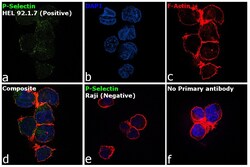
- Immunofluorescence analysis of P-Selectin was performed using HEL 92.1.7 and Raji cells. The cells were fixed with 4% paraformaldehyde for 10 minutes, permeabilized with 0.1% Triton™ X-100 for 15 minutes, and blocked with 2% BSA for 1 hour at room temperature. The cells were labeled with P-selectin Mouse Monoclonal Antibody (Product # 14-0628-80) at 5 µg/mL in 0.1% BSA and incubated overnight at 4 degree and then labeled with Goat anti-Mouse IgG (H+L) Superclonal™ Recombinant Secondary Antibody, Alexa Fluor® 488 (Product # A28175) at a dilution of 1:2000 for 45 minutes at room temperature (Panel a: green). Nuclei (Panel b: blue) were stained with ProLong™ Diamond Antifade Mountant with DAPI (Product # P36962). F-actin (Panel c: red) was stained with Rhodamine Phalloidin (Product # R415, 1:300). Panel d represents the composite image showing membrane localization of P-Selectin in HEL 92.1.7 but not in Raji (Panel e). Panel f represents control cells with no primary antibody to assess background. The images were captured at 60X magnification..

Supportive validation
- Submitted by
- Invitrogen Antibodies (provider)
- Main image

- Experimental details
- Staining of thrombin-activated human peripheral blood platelets with 0.5 µg of Mouse IgG1 kappa Isotype Control Purifed (Product # 14-4714-82) (open histogram) or 0.5 µg of Anti-Human CD62P (P-Selectin) Purified (filled histogram) followed by Anti-Mouse IgG FITC (Product # 11-4011-85). Total viable cells were used for analysis.