Explore
 Validate
 Learn
Learn17-0626-80
antibody from Invitrogen Antibodies
Targeting: SELP
CD62, CD62P, GMP140, GRMP, PADGEM, PSEL
 Flow cytometry
Antibody data
- Antibody Data
- Antigen structure
- References [15]
- Comments [0]
- Validations
- Flow cytometry [1]
- Other assay [14]
Submit
Validation data
Reference
Comment
Report error
- Product number
- 17-0626-80 - Provider product page

- Provider
- Invitrogen Antibodies
- Product name
- CD62P (P-Selectin) Monoclonal Antibody (Psel.KO2.3), APC, eBioscience™
- Antibody type
- Monoclonal
- Antigen
- Other
- Description
- Description: The Psel.KO2.3 monoclonal antibody reacts with mouse CD62P (also known as P-selectin and GMP-140), a 140-kDa cell adhesion molecule that can be induced on endothelial cells and platelets. During inflammation, this selectin mediates adhesion of monocytes and neutrophils to activated platelets, as well as leukocyte rolling on endothelial cells. The major ligand for CD62P on neutrophils is P-selectin glycoprotein ligand-1 (PSGL-1). This monoclonal antibody recognizes an epitope that is conserved across several species (e.g. mouse, human, and rat) and has not been reported to block cell adhesion. Applications Reported: This Psel.KO2.3 antibody has been reported for use in flow cytometric analysis. Applications Tested: This Psel.KO2.3 antibody has been tested by flow cytometric analysis on thrombin-activated human platelets. This can be used at less than or equal to 0.25 µg per test. A test is defined as the amount (µg) of antibody that will stain a cell sample in a final volume of 100 µL. Cell number should be determined empirically but can range from 10^5 to 10^8 cells/test. It is recommended that the antibody be carefully titrated for optimal performance in the assay of interest. Excitation: 633-647 nm; Emission: 660 nm; Laser: Red Laser. Filtration: 0.2 µm post-manufacturing filtered.
- Reactivity
- Human, Mouse
- Host
- Mouse
- Isotype
- IgG
- Antibody clone number
- Psel.KO2.3
- Vial size
- 25 µg
- Concentration
- 0.2 mg/mL
- Storage
- 4° C, store in dark, DO NOT FREEZE!
Submitted references Loss of Tet2 affects platelet function but not coagulation in mice.
Longitudinal RNA-Seq Analysis of the Repeatability of Gene Expression and Splicing in Human Platelets Identifies a Platelet SELP Splice QTL.
Cystic fibrosis transmembrane conductance regulator dysfunction in platelets drives lung hyperinflammation.
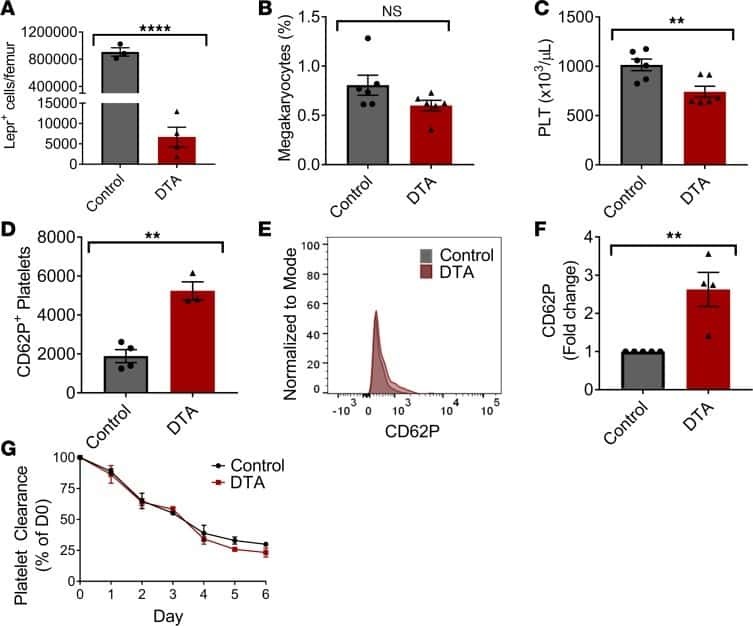
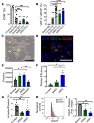
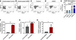
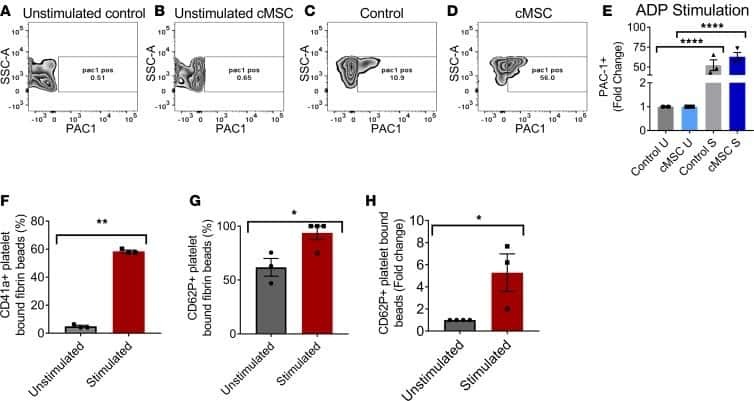
Mesenchymal stromal cells lower platelet activation and assist in platelet formation in vitro.
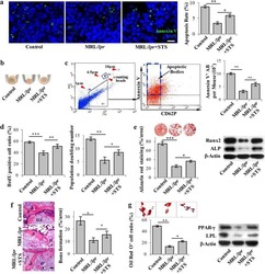
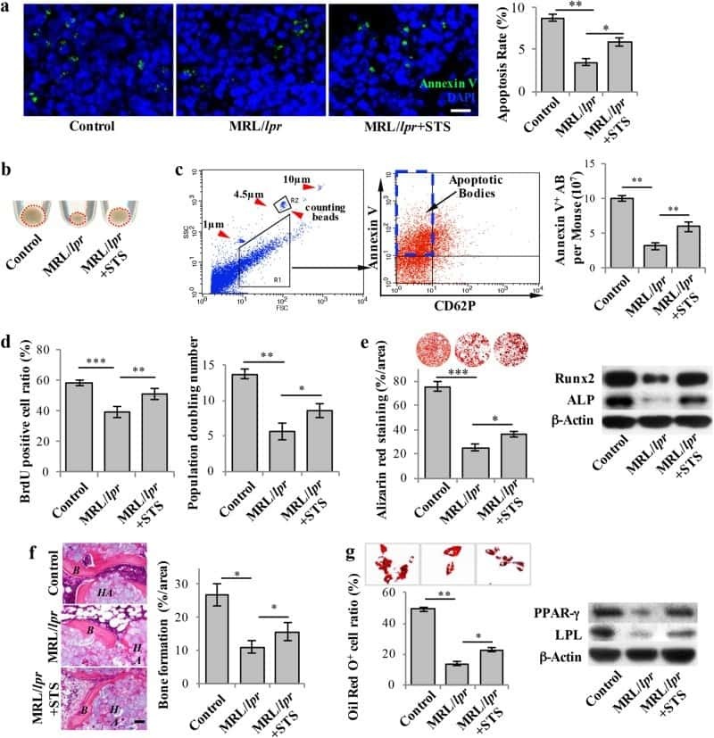
Circulating apoptotic bodies maintain mesenchymal stem cell homeostasis and ameliorate osteopenia via transferring multiple cellular factors.
Novel antibodies against GPIbα inhibit pulmonary metastasis by affecting vWF-GPIbα interaction.
Mannose-Binding Lectin Drives Platelet Inflammatory Phenotype and Vascular Damage After Cerebral Ischemia in Mice via IL (Interleukin)-1α.
The critical role of SENP1-mediated GATA2 deSUMOylation in promoting endothelial activation in graft arteriosclerosis.
Neutrophil-derived S100 calcium-binding proteins A8/A9 promote reticulated thrombocytosis and atherogenesis in diabetes.
Refrigerated platelets stored in whole blood up to 5 days adhere to thrombi formed during hemorrhagic hypotension in rats.
Platelets are relevant mediators of renal injury induced by primary endothelial lesions.
Platelet-derived HMGB1 is a critical mediator of thrombosis.
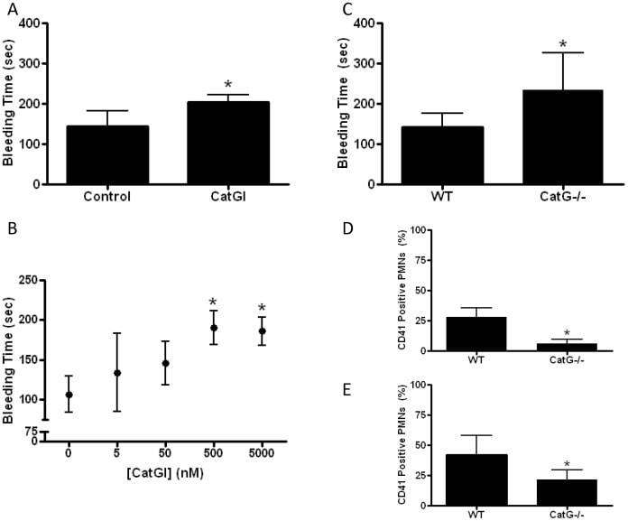
Cathepsin G-dependent modulation of platelet thrombus formation in vivo by blood neutrophils.
CD43 plays both antiadhesive and proadhesive roles in neutrophil rolling in a context-dependent manner.
Production and characterization of monoclonal antibodies against conserved epitopes of P-selectin (CD62P).
Wang B, Xia M, Chen T, Li M, Shi D, Wang X, Pang A, Zhou J, Yuan W, Chu Y
Blood science (Baltimore, Md.) 2020 Oct;2(4):129-136
Blood science (Baltimore, Md.) 2020 Oct;2(4):129-136
Longitudinal RNA-Seq Analysis of the Repeatability of Gene Expression and Splicing in Human Platelets Identifies a Platelet SELP Splice QTL.
Rondina MT, Voora D, Simon LM, Schwertz H, Harper JF, Lee O, Bhatlekar SC, Li Q, Eustes AS, Montenont E, Campbell RA, Tolley ND, Kosaka Y, Weyrich AS, Bray PF, Rowley JW
Circulation research 2020 Feb 14;126(4):501-516
Circulation research 2020 Feb 14;126(4):501-516
Cystic fibrosis transmembrane conductance regulator dysfunction in platelets drives lung hyperinflammation.
Ortiz-Muñoz G, Yu MA, Lefrançais E, Mallavia B, Valet C, Tian JJ, Ranucci S, Wang KM, Liu Z, Kwaan N, Dawson D, Kleinhenz ME, Khasawneh FT, Haggie PM, Verkman AS, Looney MR
The Journal of clinical investigation 2020 Apr 1;130(4):2041-2053
The Journal of clinical investigation 2020 Apr 1;130(4):2041-2053
Mesenchymal stromal cells lower platelet activation and assist in platelet formation in vitro.
Mendelson A, Strat AN, Bao W, Rosston P, Fallon G, Ohrn S, Zhong H, Lobo C, An X, Yazdanbakhsh K
JCI insight 2019 Aug 22;4(16)
JCI insight 2019 Aug 22;4(16)
Circulating apoptotic bodies maintain mesenchymal stem cell homeostasis and ameliorate osteopenia via transferring multiple cellular factors.
Liu D, Kou X, Chen C, Liu S, Liu Y, Yu W, Yu T, Yang R, Wang R, Zhou Y, Shi S
Cell research 2018 Sep;28(9):918-933
Cell research 2018 Sep;28(9):918-933
Novel antibodies against GPIbα inhibit pulmonary metastasis by affecting vWF-GPIbα interaction.
Qi Y, Chen W, Liang X, Xu K, Gu X, Wu F, Fan X, Ren S, Liu J, Zhang J, Li R, Liu J, Liang X
Journal of hematology & oncology 2018 Sep 17;11(1):117
Journal of hematology & oncology 2018 Sep 17;11(1):117
Mannose-Binding Lectin Drives Platelet Inflammatory Phenotype and Vascular Damage After Cerebral Ischemia in Mice via IL (Interleukin)-1α.
Orsini F, Fumagalli S, Császár E, Tóth K, De Blasio D, Zangari R, Lénárt N, Dénes Á, De Simoni MG
Arteriosclerosis, thrombosis, and vascular biology 2018 Nov;38(11):2678-2690
Arteriosclerosis, thrombosis, and vascular biology 2018 Nov;38(11):2678-2690
The critical role of SENP1-mediated GATA2 deSUMOylation in promoting endothelial activation in graft arteriosclerosis.
Qiu C, Wang Y, Zhao H, Qin L, Shi Y, Zhu X, Song L, Zhou X, Chen J, Zhou H, Zhang H, Tellides G, Min W, Yu L
Nature communications 2017 Jun 1;8:15426
Nature communications 2017 Jun 1;8:15426
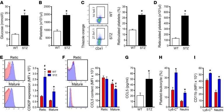
Neutrophil-derived S100 calcium-binding proteins A8/A9 promote reticulated thrombocytosis and atherogenesis in diabetes.
Kraakman MJ, Lee MK, Al-Sharea A, Dragoljevic D, Barrett TJ, Montenont E, Basu D, Heywood S, Kammoun HL, Flynn M, Whillas A, Hanssen NM, Febbraio MA, Westein E, Fisher EA, Chin-Dusting J, Cooper ME, Berger JS, Goldberg IJ, Nagareddy PR, Murphy AJ
The Journal of clinical investigation 2017 Jun 1;127(6):2133-2147
The Journal of clinical investigation 2017 Jun 1;127(6):2133-2147
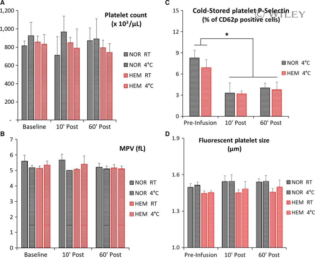
Refrigerated platelets stored in whole blood up to 5 days adhere to thrombi formed during hemorrhagic hypotension in rats.
Torres Filho IP, Torres LN, Valdez C, Salgado C, Cap AP, Dubick MA
Journal of thrombosis and haemostasis : JTH 2017 Jan;15(1):163-175
Journal of thrombosis and haemostasis : JTH 2017 Jan;15(1):163-175
Platelets are relevant mediators of renal injury induced by primary endothelial lesions.
Schwarzenberger C, Sradnick J, Lerea KM, Goligorsky MS, Nieswandt B, Hugo CP, Hohenstein B
American journal of physiology. Renal physiology 2015 Jun 1;308(11):F1238-46
American journal of physiology. Renal physiology 2015 Jun 1;308(11):F1238-46
Platelet-derived HMGB1 is a critical mediator of thrombosis.
Vogel S, Bodenstein R, Chen Q, Feil S, Feil R, Rheinlaender J, Schäffer TE, Bohn E, Frick JS, Borst O, Münzer P, Walker B, Markel J, Csanyi G, Pagano PJ, Loughran P, Jessup ME, Watkins SC, Bullock GC, Sperry JL, Zuckerbraun BS, Billiar TR, Lotze MT, Gawaz M, Neal MD
The Journal of clinical investigation 2015 Dec;125(12):4638-54
The Journal of clinical investigation 2015 Dec;125(12):4638-54
Cathepsin G-dependent modulation of platelet thrombus formation in vivo by blood neutrophils.
Faraday N, Schunke K, Saleem S, Fu J, Wang B, Zhang J, Morrell C, Dore S
PloS one 2013;8(8):e71447
PloS one 2013;8(8):e71447
CD43 plays both antiadhesive and proadhesive roles in neutrophil rolling in a context-dependent manner.
Matsumoto M, Shigeta A, Miyasaka M, Hirata T
Journal of immunology (Baltimore, Md. : 1950) 2008 Sep 1;181(5):3628-35
Journal of immunology (Baltimore, Md. : 1950) 2008 Sep 1;181(5):3628-35
Production and characterization of monoclonal antibodies against conserved epitopes of P-selectin (CD62P).
Massaguer A, Engel P, Pérez-del-Pulgar S, Bosch J, Pizcueta P
Tissue antigens 2000 Aug;56(2):117-28
Tissue antigens 2000 Aug;56(2):117-28
No comments: Submit comment
Supportive validation
- Submitted by
- Invitrogen Antibodies (provider)
- Main image
- Experimental details
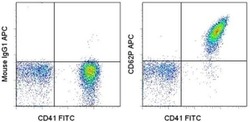
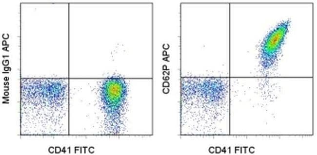
- Staining of thrombin-activated human platelets with Anti-Human CD41a FITC (Product # 11-0419-42) and 0.125 µg of Mouse IgG1 K Isotype Control APC (Product # 17-4714-81) (left) or 0.125 µg of Anti-Human/Mouse CD62P (P-selectin) APC (right). Total viable cells were used for analysis.
Supportive validation
- Submitted by
- Invitrogen Antibodies (provider)
- Main image

- Experimental details
- NULL
- Submitted by
- Invitrogen Antibodies (provider)
- Main image

- Experimental details
- NULL
- Submitted by
- Invitrogen Antibodies (provider)
- Main image
- Experimental details
- NULL
- Submitted by
- Invitrogen Antibodies (provider)
- Main image
- Experimental details
- NULL
- Submitted by
- Invitrogen Antibodies (provider)
- Main image
- Experimental details
- NULL

- Submitted by
- Invitrogen Antibodies (provider)
- Main image
- Experimental details
- NULL
- Submitted by
- Invitrogen Antibodies (provider)
- Main image
- Experimental details
- NULL
- Submitted by
- Invitrogen Antibodies (provider)
- Main image
- Experimental details
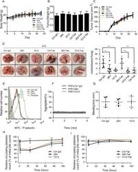
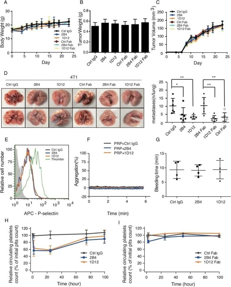
- Fig. 4 2B4 and 1D12 inhibit spontaneous metastasis but have no effect on platelet activation and hemostatic function. Effect of 2B4 (50 mug/mouse), 1D12 (50 mug/mouse), and their Fabs (50 mug/mouse) on ( a ) mice weight, ( b ) tumor weight, ( c ) tumor volume, and ( d ) pulmonary metastasis on spontaneous metastasis of 4T1 ( n = 6 mice in each group). Metastasis was analyzed 3 weeks after injection of tumor cells. Representative examples of the lungs (one of each group) with metastatic foci were depicted. Average number of lung metastasis in each of the groups was shown in right graphs (+- SD, P value is indicated; * P < 0.05; ** P < 0.01; *** P < 0.001). e 2B4 and 1D12 did not affect platelet activation. Increased expression of APC-conjugated P-selectin indicated the degree of platelet activation. Washed platelets were treated with 10 mug/ml purified 2B4 (blue), 1D12 (orange), negative control (rat IgG, gray), or 0.05 U/ml thrombin (green) as positive control, then probed with APC-conjugated anti-P-selectin Ab. The florescence intensity was detected by flow cytometry. f 2B4 and 1D12 did not induce platelet aggregation. The aggregation of PRP pretreated with 10 mug/ml 2B4 (blue) and 10 mug/ml 1D12 (orange) was detected. Negative control was in the form of rat IgG. g Bleeding time did not prolonged 2 h after the injection of intact 2B4 or 1D12 (50 mug/mouse) ( n = 4 mice in each group). h Platelet survival curves for mice injected with 2B4 (50 mug/mouse), 1D12 (
- Submitted by
- Invitrogen Antibodies (provider)
- Main image
- Experimental details
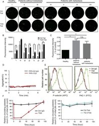
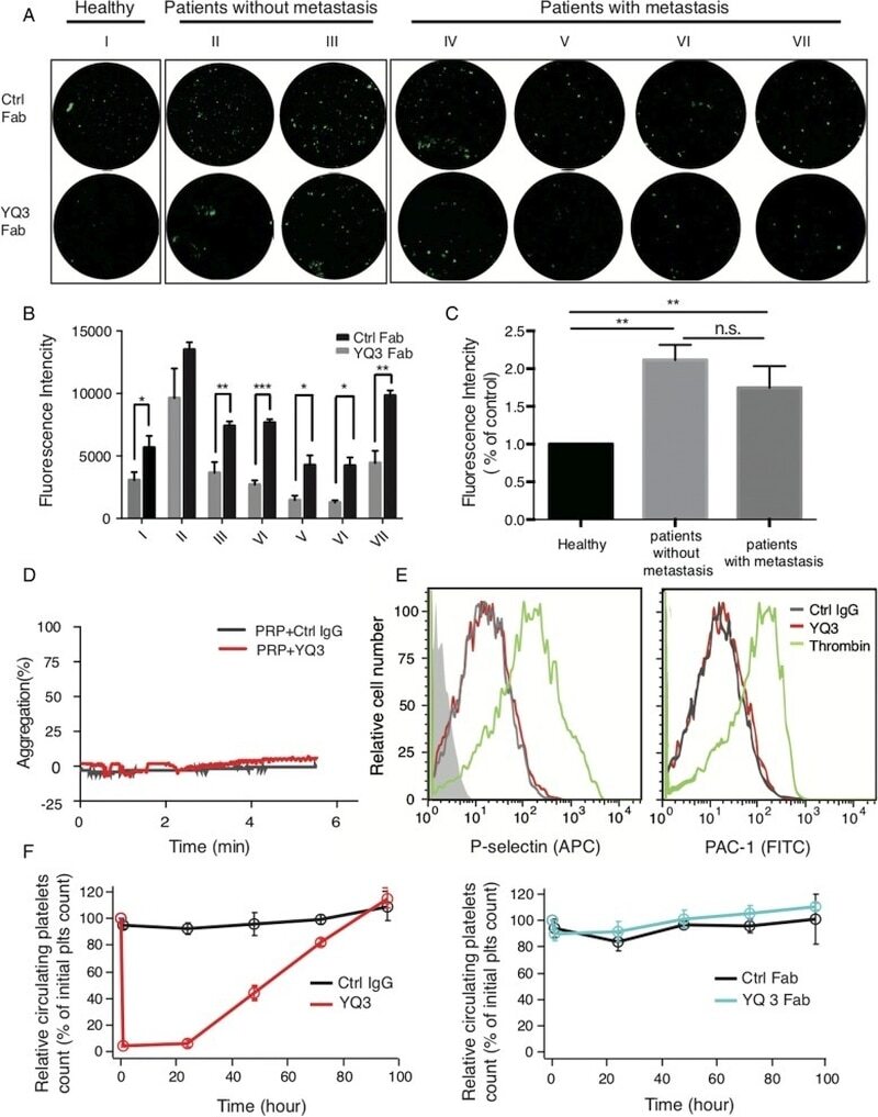
- Fig. 7 YQ3 inhibits the adhesion between patients' platelets and tumor cells without accelerating platelet clearance. a , b YQ3 inhibited adhesion of A549 lung cancer cells to patients' platelets. a The adhesion of A549 to patients' platelets pretreated with 10 mug/ml YQ3 Fab as observed under fluorescence microscope. I: healthy person; II/III: patients without metastasis; IV/V/VI/VII: patients with metastasis. The quantitative analysis of adhesion was shown in b , P value is indicated; * P < 0.05; ** P < 0.01; *** P < 0.001. c Expression of GPIbalpha on platelets that were from healthy controls, patients without metastasis, or patients with metastasis. Fluorescence intensity was detected by flow cytometry. ** P < 0.01, n.s., no signifance. d YQ3 did not induce platelet aggregation. The aggregation of PRP pretreated with 10 mug/ml YQ3 was detected. Negative control was in the form of normal mouse IgG. e YQ3 did not affect platelet activation. Increased expression of P-selectin and PAC-1 indicated the degree of platelet activation. Washed human platelets were treated with 10 mug/ml purified YQ3 (red) and negative control (mouse IgG, gray) or 0.05 U/ml thrombin (green) as positive control, then probed with APC-conjugated P-selectin Ab and FITC-conjugated APC-1 Ab. The fluorescence intensity was detected by flow cytometry. f Platelet survival curves for hTg mice injected with YQ3 (15 mug/mouse), YQ3 Fab (15 mug/mouse), or negative control (mouse IgG or Fab, 1
- Submitted by
- Invitrogen Antibodies (provider)
- Main image
- Experimental details
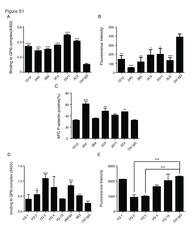
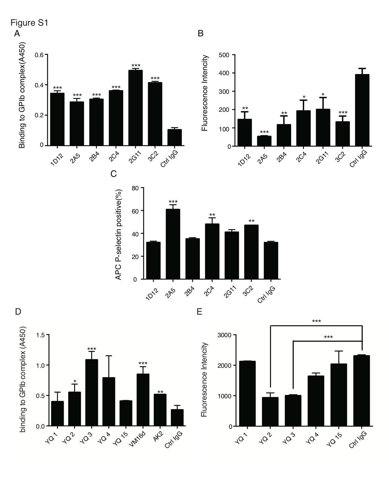
- Additional file 2: Figure S1. Screening of six rat anti-mouse GPIbalpha antibodies and five mouse anti-human GPIbalpha antibodies. (A) The quantitative analysis of adhesion of LLC cells with BCECF-labeled mouse platelets in the presence of various antibodies was measured of fluorescent intensity under fluorescence plate reader. (B) Effect of antibodies on platelet activation was detected by flow cytometry. Washed platelets were treated with hybridoma supernatant and negative control (RPMI-1640 fetal bovine culture medium with rat IgG) and then probed with APC-conjugated anti-P-selectin Ab. (C) Purified of 2B4 and 1D12 and its Fab fragments were run in 10% Bis-Tris SDS gel electrophoresis under reducing (r.) and nonreducing (n.r.) conditions. Molecular weight marker (M) was shown and labeled in kDa. (D) The quantitative analysis of adhesion of HCT116 cells with BCECF-labeled human platelets in the presence of various antibodies was measured of fluorescent intensity under fluorescence plate reader. (E) Purified of YQ3 and its Fab fragment were run in 10% Bis-Tris SDS gel electrophoresis under reducing (r.) and nonreducing (n.r.) conditions. Molecular weight marker (M) was shown and labeled in kDa on the left. P value is indicated; * P < 0.05; ** P < 0.01; *** P < 0.001. Each figure is a representative of three independent experiments. (TIFF 8219 kb)
- Submitted by
- Invitrogen Antibodies (provider)
- Main image

- Experimental details
- Figure 2 Cathepsin G promotes hemostasis after tissue injury in the mouse in vivo. A . Tail bleeding time was determined in mice after intravenous injection of vehicle (control) or cathepsin G inhibitor I (CatGI, 500 nM final); N = 12 each group, *P
- Submitted by
- Invitrogen Antibodies (provider)
- Main image
- Experimental details
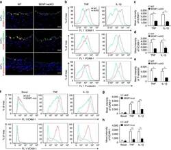
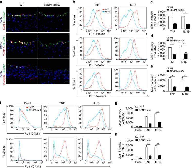
- Figure 3 Loss of endothelial SENP1 inhibits EC activation. ( a ) Grafts from WT or SENP1-ecKO mice were harvested 3 days post-transplantation. The induction of endothelial adhesion molecules was demonstrated by immunofluorescence staining of ICAM-1, VCAM-1, or P-selectin and PECAM-1 with DAPI labelling of the nuclei. Bar represents 50 mum. ( b - e ) Attenuated induction of adhesion molecules in SENP1-ecKO MAECs. Flow cytometry analysis of ICAM-1, VCAM-1 and P-selectin in MAECs isolated from WT or SENP1-ecKO mice after TNF or IL-1beta treatment. Representative histograms are shown in ( b ) with the quantification of mean intensity in ( c - e ). ( f - h ) Overexpression of the catalytically inactive form of SENP1 (SENP1-Mut) inhibits the induction of adhesion molecules in HUVECs. HUVECs were infected by Ad-SENP1-Mut or vector control (Ad-LacZ) for 24 h, treated with pro-inflammatory cytokines and analysed by flow cytometry in the same way as MAECs. Representative histograms of ICAM-1 and VCAM-1 are shown in ( f ) with the quantification of mean intensity in ( g , h ). Data are presented as the mean+-s.e.m. from at least three independent experiments. * P
- Submitted by
- Invitrogen Antibodies (provider)
- Main image

- Experimental details
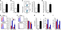
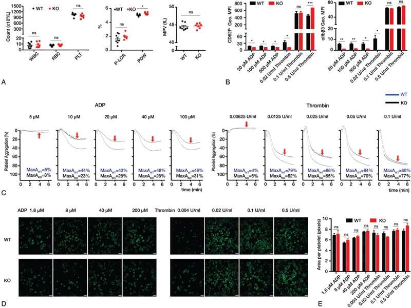
- Figure 1. Decreased functions of platelets in Tet2 -deficient mice. (A) The blood routine results of WT and Tet2 KO mice showed no significant differences in their WBC, RBC, and PLT counts (left panel), and in P-LCR (middle panel) and MPV (right panel), ( Tet2 KO, n&hairsp=&hairsp7; WT, n&hairsp=&hairsp7). Only PDW (middle panel) was increased moderately. (B) Flow cytometry was used to evaluate PB platelet activation with agonists in Tet2 KO (n&hairsp=&hairsp4) and WT (n&hairsp=&hairsp5) mice. CD62P (left) and &agrIIb&bgr3 (right) were the surface markers used for detection of platelet activation. (C) Aggregation assays of washed platelets (2&hairspx&hairsp10 8 &solml) were performed with Chrono-log Model 700 Whole Blood&solOptical Lumi-Aggregometers. When stimulated by ADP (right) and medium concentrations of thrombin (left), the aggregation of washed platelets of Tet2 KO mice was lower than WT mice. Black curve and red arrow represent Tet2 KO platelets while blue one represents WT platelets. MaxA, maximum aggregation (&percnt). (D and E) Phalloidin-FITC staining of washed platelets (3&hairspx&hairsp10 7 &solml) with or without agonists from PB of Tet2 KO and WT mice. The representative images were displayed (D). Scale bar: 6&hairsp&mgrm. Quantification of the area of each platelet (E, n&hairsp=&hairsp3). Data was processing with Volocity software. MPV, mean platelet volume; PB, peripheral blood; PDW, platelet distribution width; P-LCR, platelet-larger cell ratio; PLT, plate
- Submitted by
- Invitrogen Antibodies (provider)
- Main image

- Experimental details
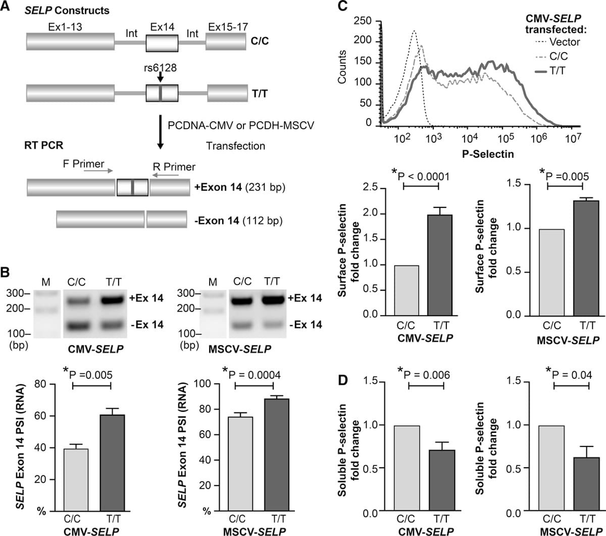
- Figure 7. rs6128 directly regulates exon 14 skipping in SELP and alters the ratio of surface to soluble P-selectin protein expression. A , Schematic of mini-gene constructs of SELP that include the open reading frame of SELP , and the introns flanking exon 14. The C/C and T/T constructs vary by a single nucleotide at rs6128. Constructs were cloned into vectors with 2 different promoters (cytomegalovirus [CMV] or murine stem cell virus [MSCV]). After transfection into HEK (human embryonic kidney) 293 cells, the introns are spliced out, and exon 14 is variably spliced out (skipped). The extent of exon 14 skipping is measured by PCR via exon 14 flanking primers that generate 2 polymerase chain reaction (PCR) products of different sizes. B , Real-time PCR analysis of SELP exon 14 skipping following transfection of HEK 293 cells with rs6128 C/C or T/T vectors. Shown is a representative result from 5 independent experiments. Below are bar graphs and SE summary of PSI calculated according to densitometry analysis of the exon 14 inclusion band (upper band) divided by the sum of the upper and lower bands (total).*paired t test, n=5 independent experiments. C , Flow cytometry analysis of P-selectin surface expression following transfection of HEK 293 cells with rs6128 C/C or T/T vectors. Top , Representative histogram overlay of P-selectin surface expression 24 h after transfection with CMV promoter empty vector, rs6128 C/C, or T/