Explore
 Validate
 Learn
LearnA01241-1
antibody from Boster Biological Technology
Targeting: SELP
CD62, CD62P, GMP140, GRMP, PADGEM, PSEL
 Western blot
 ELISA
ELISAAntibody data
- Antibody Data
- Antigen structure
- References [3]
- Comments [0]
- Validations
- Western blot [1]
Submit
Validation data
Reference
Comment
Report error
- Product number
- A01241-1 - Provider product page

- Provider
- Boster Biological Technology
- Product name
- Anti-CD62P/Selp Antibody Picoband™
- Antibody type
- Polyclonal
- Description
- Rabbit IgG polyclonal antibody for CD62P detection. Tested with WB, IHC-F, ICC, FCM, Direct ELISA in Mouse;Rat.
- Reactivity
- Mouse, Rat
- Host
- Rabbit
- Vial size
- 100μg/vial
- Concentration
- 0.5-1mg/ml, actual concentration vary by lot. Use suggested dilution ratio to decide dilution procedure.
- Storage
- At -20°C for one year. After reconstitution, at 4°C for one month. It can also be aliquoted and stored frozen at -20°C for a longer time. Avoid repeated freezing and thawing.
- Handling
- Add 0.2ml of distilled water will yield a concentration of 500ug/ml.
Submitted references Gynura segetum induces hepatic sinusoidal obstruction syndrome in mice by impairing autophagy.
Irisin Pretreatment Protects Kidneys against Acute Kidney Injury Induced by Ischemia/Reperfusion via Upregulating the Expression of Uncoupling Protein 2.
Fabrication of biomolecule-PEG micropattern on titanium surface and its effects on platelet adhesion.
Zhang H, Jia S, Jin L, Mb JY, Shen Z, Wu J, Yao X, Chen D, Zhang C, Yu S, Zhu N, Jin L, Yao X
Acta cirurgica brasileira 2022;36(11):e361104
Acta cirurgica brasileira 2022;36(11):e361104
Irisin Pretreatment Protects Kidneys against Acute Kidney Injury Induced by Ischemia/Reperfusion via Upregulating the Expression of Uncoupling Protein 2.
Zhang R, Ji J, Zhou X, Li R
BioMed research international 2020;2020:6537371
BioMed research international 2020;2020:6537371
Fabrication of biomolecule-PEG micropattern on titanium surface and its effects on platelet adhesion.
Zhang F, Li G, Yang P, Qin W, Li C, Huang N
Colloids and surfaces. B, Biointerfaces 2013 Feb 1;102:457-65
Colloids and surfaces. B, Biointerfaces 2013 Feb 1;102:457-65
No comments: Submit comment
Supportive validation
- Submitted by
- Boster Biological Technology (provider)
- Main image
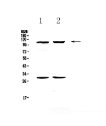
- Experimental details
- Western blot analysis of CD62P using anti-CD62P antibody (A01241-1). Electrophoresis was performed on a 5-20% SDS-PAGE gel at 70V (Stacking gel) / 90V (Resolving gel) for 2-3 hours. The sample well of each lane was loaded with 50ug of sample under reducing conditions. Lane 1: mouse heart tissue lysates,Lane 2: rat heart tissue lysates. After Electrophoresis, proteins were transferred to a Nitrocellulose membrane at 150mA for 50-90 minutes. Blocked the membrane with 5% Non-fat Milk/ TBS for 1.5 hour at RT. The membrane was incubated with rabbit anti-CD62P antigen affinity purified polyclonal antibody (Catalog # A01241-1) at 0.5 μg/mL overnight at 4°C, then washed with TBS-0.1%Tween 3 times with 5 minutes each and probed with a goat anti-rabbit IgG-HRP secondary antibody at a dilution of 1:10000 for 1.5 hour at RT. The signal is developed using an Enhanced Chemiluminescent detection (ECL) kit (Catalog # EK1002) with Tanon 5200 system. A specific band was detected for CD62P at approximately 110KD. The expected band size for CD62P is at 91KD.
- Additional image