 Explore
 Validate
 Learn
Learn Western blot
 Immunohistochemistry
Antibody data
- Antibody Data
- Antigen structure
- References [3]
- Comments [0]
- Validations
- Western blot [1]
- Flow cytometry [3]
Submit
Validation data
Reference
Comment
Report error
- Product number
- MA1-19028 - Provider product page

- Provider
- Invitrogen Antibodies
- Product name
- ICAM-1 Monoclonal Antibody (1H4)
- Antibody type
- Monoclonal
- Antigen
- Other
- Description
- For IHC (P), prolonged fixation in buffered formalin can destroy the epitope. High temperature antigen unmasking technique is required.
- Reactivity
- Human
- Host
- Mouse
- Isotype
- IgG
- Antibody clone number
- 1H4
- Vial size
- 100 µg
- Concentration
- 1 mg/mL
- Storage
- 4° C, do not freeze
Submitted references Interaction between adipose tissue stromal cells and gastric cancer cells in vitro.
Adipose tissue explants and MDCK cells reciprocally regulate their morphogenesis in coculture.
A new organotypic culture of adipose tissue fragments maintains viable mature adipocytes for a long term, together with development of immature adipocytes and mesenchymal stem cell-like cells.
Nomoto-Kojima N, Aoki S, Uchihashi K, Matsunobu A, Koike E, Ootani A, Yonemitsu N, Fujimoto K, Toda S
Cell and tissue research 2011 May;344(2):287-98
Cell and tissue research 2011 May;344(2):287-98
Adipose tissue explants and MDCK cells reciprocally regulate their morphogenesis in coculture.
Udo K, Aoki S, Uchihashi K, Kawasaki M, Matsunobu A, Tokuda Y, Ootani A, Toda S, Uozumi J
Kidney international 2010 Jul;78(1):60-8
Kidney international 2010 Jul;78(1):60-8
A new organotypic culture of adipose tissue fragments maintains viable mature adipocytes for a long term, together with development of immature adipocytes and mesenchymal stem cell-like cells.
Sonoda E, Aoki S, Uchihashi K, Soejima H, Kanaji S, Izuhara K, Satoh S, Fujitani N, Sugihara H, Toda S
Endocrinology 2008 Oct;149(10):4794-8
Endocrinology 2008 Oct;149(10):4794-8
No comments: Submit comment
Supportive validation
- Submitted by
- Invitrogen Antibodies (provider)
- Main image
- Experimental details
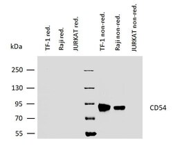
- Western blotting analysis of human CD54 using mouse monoclonal antibody 1H4 on lysates of TF-1 and Raji cells, as well as of JURKAT cells (negative control) under reducing and non-reducing conditions. Nitrocellulose membrane was probed with 2µg/mL of mouse anti-CD54 Monoclonal antibody (Product # MA1-19028) followed by IRDye800-conjugated anti-mouse secondary antibody. A specific band was detected for CD54 at approximately 88kDa.
Supportive validation
- Submitted by
- Invitrogen Antibodies (provider)
- Main image
- Experimental details
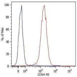
- Flow cytometry surface staining of U937 human histiocytic lymphoma cell line with anti-human CD54 monoclonal antibody (Product # MA1-19028). Total viable cells were used for analysis.

- Submitted by
- Invitrogen Antibodies (provider)
- Main image
- Experimental details
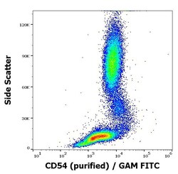
- Flow cytometry surface staining pattern of human peripheral whole blood stained using anti-human CD54 (1H4) purified Monoclonal antibody (Product # MA1-19028) (concentration in sample 3 µg/mL, GAM FITC).

- Submitted by
- Invitrogen Antibodies (provider)
- Main image

- Experimental details
- Separation of human monocytes (red-filled) from human lymphocytes (black-dashed) in flow cytometry analysis (surface staining) of peripheral whole blood stained using anti-human CD54 (1H4) purified Monoclonal antibody (Product # MA1-19028) (concentration in sample 3 µg/mL, GAM FITC).
