Explore
 Validate
 Learn
Learn Western blot
 Immunocytochemistry
 Immunohistochemistry
Antibody data
- Antibody Data
- Antigen structure
- References [11]
- Comments [0]
- Validations
- Immunocytochemistry [5]
- Flow cytometry [2]
- Other assay [1]
Submit
Validation data
Reference
Comment
Report error
- Product number
- MA5-13021 - Provider product page

- Provider
- Invitrogen Antibodies
- Product name
- ICAM-1 Monoclonal Antibody (W-CAM-1)
- Antibody type
- Monoclonal
- Antigen
- Other
- Description
- MA5-13021 targets CD54 in WB, FACS, FN, IF, and IHC (F) applications and shows reactivity with Human samples. The MA5-13021 immunogen is raji Burkitt lymphoma cells.
- Reactivity
- Human
- Host
- Mouse
- Isotype
- IgG
- Antibody clone number
- W-CAM-1
- Vial size
- 500 μL
- Concentration
- 0.2 mg/mL
- Storage
- 4°C
Submitted references Expanding the clinical and metabolic phenotype of DPM2 deficient congenital disorders of glycosylation.
Vγ9Vδ2 T Cells Activation Through Phosphoantigens Can Be Impaired by a RHOB Rerouting in Lung Cancer.
Transdifferentiated Human Vascular Smooth Muscle Cells are a New Potential Cell Source for Endothelial Regeneration.
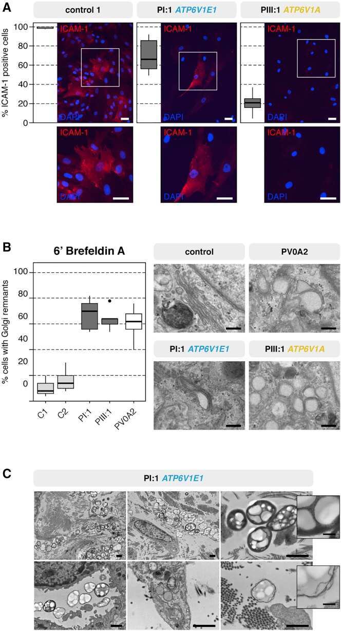
Mutations in ATP6V1E1 or ATP6V1A Cause Autosomal-Recessive Cutis Laxa.
Galactose Supplementation in Patients With TMEM165-CDG Rescues the Glycosylation Defects.
Exosomes derived from Epstein-Barr virus-infected cells are internalized via caveola-dependent endocytosis and promote phenotypic modulation in target cells.
Intercellular adhesion molecule (ICAM)-1 and vascular cell adhesion molecule (VCAM)-1 at the early stages of atherosclerosis in a rat model.
Interaction between adipose tissue stromal cells and gastric cancer cells in vitro.
Adipose tissue explants and MDCK cells reciprocally regulate their morphogenesis in coculture.
A new organotypic culture of adipose tissue fragments maintains viable mature adipocytes for a long term, together with development of immature adipocytes and mesenchymal stem cell-like cells.
Regulation of expression of a human intercellular adhesion molecule (ICAM-1) during lymphohematopoietic differentiation.
Radenkovic S, Fitzpatrick-Schmidt T, Byeon SK, Madugundu AK, Saraswat M, Lichty A, Wong SYW, McGee S, Kubiak K, Ligezka A, Ranatunga W, Zhang Y, Wood T, Friez MJ, Clarkson K, Pandey A, Jones JR, Morava E
Molecular genetics and metabolism 2021 Jan;132(1):27-37
Molecular genetics and metabolism 2021 Jan;132(1):27-37
Vγ9Vδ2 T Cells Activation Through Phosphoantigens Can Be Impaired by a RHOB Rerouting in Lung Cancer.
Laplagne C, Meddour S, Figarol S, Michelas M, Calvayrac O, Favre G, Laurent C, Fournié JJ, Cabantous S, Poupot M
Frontiers in immunology 2020;11:1396
Frontiers in immunology 2020;11:1396
Transdifferentiated Human Vascular Smooth Muscle Cells are a New Potential Cell Source for Endothelial Regeneration.
Hong X, Margariti A, Le Bras A, Jacquet L, Kong W, Hu Y, Xu Q
Scientific reports 2017 Jul 17;7(1):5590
Scientific reports 2017 Jul 17;7(1):5590
Mutations in ATP6V1E1 or ATP6V1A Cause Autosomal-Recessive Cutis Laxa.
Van Damme T, Gardeitchik T, Mohamed M, Guerrero-Castillo S, Freisinger P, Guillemyn B, Kariminejad A, Dalloyaux D, van Kraaij S, Lefeber DJ, Syx D, Steyaert W, De Rycke R, Hoischen A, Kamsteeg EJ, Wong SY, van Scherpenzeel M, Jamali P, Brandt U, Nijtmans L, Korenke GC, Chung BHY, Mak CCY, Hausser I, Kornak U, Fischer-Zirnsak B, Strom TM, Meitinger T, Alanay Y, Utine GE, Leung PKC, Ghaderi-Sohi S, Coucke P, Symoens S, De Paepe A, Thiel C, Haack TB, Malfait F, Morava E, Callewaert B, Wevers RA
American journal of human genetics 2017 Feb 2;100(2):216-227
American journal of human genetics 2017 Feb 2;100(2):216-227
Galactose Supplementation in Patients With TMEM165-CDG Rescues the Glycosylation Defects.
Morelle W, Potelle S, Witters P, Wong S, Climer L, Lupashin V, Matthijs G, Gadomski T, Jaeken J, Cassiman D, Morava E, Foulquier F
The Journal of clinical endocrinology and metabolism 2017 Apr 1;102(4):1375-1386
The Journal of clinical endocrinology and metabolism 2017 Apr 1;102(4):1375-1386
Exosomes derived from Epstein-Barr virus-infected cells are internalized via caveola-dependent endocytosis and promote phenotypic modulation in target cells.
Nanbo A, Kawanishi E, Yoshida R, Yoshiyama H
Journal of virology 2013 Sep;87(18):10334-47
Journal of virology 2013 Sep;87(18):10334-47
Intercellular adhesion molecule (ICAM)-1 and vascular cell adhesion molecule (VCAM)-1 at the early stages of atherosclerosis in a rat model.
Fotis L, Agrogiannis G, Vlachos IS, Pantopoulou A, Margoni A, Kostaki M, Verikokos C, Tzivras D, Mikhailidis DP, Perrea D
In vivo (Athens, Greece) 2012 Mar-Apr;26(2):243-50
In vivo (Athens, Greece) 2012 Mar-Apr;26(2):243-50
Interaction between adipose tissue stromal cells and gastric cancer cells in vitro.
Nomoto-Kojima N, Aoki S, Uchihashi K, Matsunobu A, Koike E, Ootani A, Yonemitsu N, Fujimoto K, Toda S
Cell and tissue research 2011 May;344(2):287-98
Cell and tissue research 2011 May;344(2):287-98
Adipose tissue explants and MDCK cells reciprocally regulate their morphogenesis in coculture.
Udo K, Aoki S, Uchihashi K, Kawasaki M, Matsunobu A, Tokuda Y, Ootani A, Toda S, Uozumi J
Kidney international 2010 Jul;78(1):60-8
Kidney international 2010 Jul;78(1):60-8
A new organotypic culture of adipose tissue fragments maintains viable mature adipocytes for a long term, together with development of immature adipocytes and mesenchymal stem cell-like cells.
Sonoda E, Aoki S, Uchihashi K, Soejima H, Kanaji S, Izuhara K, Satoh S, Fujitani N, Sugihara H, Toda S
Endocrinology 2008 Oct;149(10):4794-8
Endocrinology 2008 Oct;149(10):4794-8
Regulation of expression of a human intercellular adhesion molecule (ICAM-1) during lymphohematopoietic differentiation.
Boyd AW, Dunn SM, Fecondo JV, Culvenor JG, Dührsen U, Burns GF, Wawryk SO
Blood 1989 May 15;73(7):1896-903
Blood 1989 May 15;73(7):1896-903
No comments: Submit comment
Supportive validation
- Submitted by
- Invitrogen Antibodies (provider)
- Main image
- Experimental details
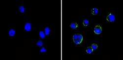
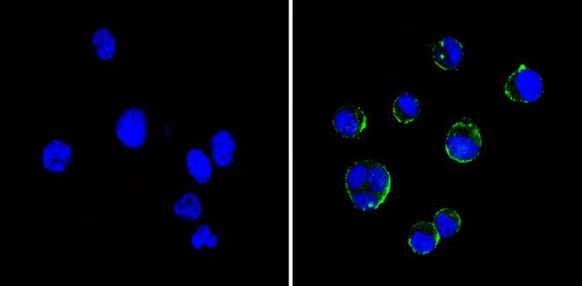
- Immunofluorescent analysis of CD54/ICAM-1 (green) showing staining in the membrane of Raji cells (right) compared to a negative control without primary antibody (left). Formalin-fixed cells were permeabilized with 0.1% Triton X-100 in TBS for 5-10 minutes and blocked with 3% BSA-PBS for 30 minutes at room temperature. Cells were probed with a CD54 monoclonal antibody (Product # MA5-13021) in 3% BSA-PBS at a dilution of 1:20 and incubated overnight at 4ºC in a humidified chamber. Cells were washed with PBST and incubated with a DyLight-conjugated secondary antibody in PBS at room temperature in the dark. F-actin (red) was stained with a fluorescent red phalloidin and nuclei (blue) were stained with Hoechst or DAPI. Images were taken at a magnification of 60x.

- Submitted by
- Invitrogen Antibodies (provider)
- Main image

- Experimental details
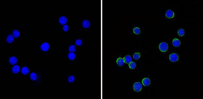
- Immunofluorescent analysis of CD54/ICAM-1 (green) showing staining in the membrane of Ramos cells (right) compared to a negative control without primary antibody (left). Formalin-fixed cells were permeabilized with 0.1% Triton X-100 in TBS for 5-10 minutes and blocked with 3% BSA-PBS for 30 minutes at room temperature. Cells were probed with a CD54 monoclonal antibody (Product # MA5-13021) in 3% BSA-PBS at a dilution of 1:20 and incubated overnight at 4ºC in a humidified chamber. Cells were washed with PBST and incubated with a DyLight-conjugated secondary antibody in PBS at room temperature in the dark. F-actin (red) was stained with a fluorescent red phalloidin and nuclei (blue) were stained with Hoechst or DAPI. Images were taken at a magnification of 60x.
- Submitted by
- Invitrogen Antibodies (provider)
- Main image

- Experimental details
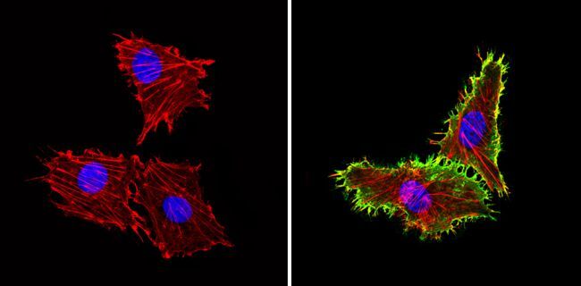
- Immunofluorescent analysis of CD54/ICAM-1 (green) showing staining in the membrane of HUVEC cells (right) compared to a negative control without primary antibody (left). Formalin-fixed cells were permeabilized with 0.1% Triton X-100 in TBS for 5-10 minutes and blocked with 3% BSA-PBS for 30 minutes at room temperature. Cells were probed with a CD54 monoclonal antibody (Product # MA5-13021) in 3% BSA-PBS at a dilution of 1:20 and incubated overnight at 4ºC in a humidified chamber. Cells were washed with PBST and incubated with a DyLight-conjugated secondary antibody in PBS at room temperature in the dark. F-actin (red) was stained with a fluorescent red phalloidin and nuclei (blue) were stained with Hoechst or DAPI. Images were taken at a magnification of 60x.
- Submitted by
- Invitrogen Antibodies (provider)
- Main image

- Experimental details
- Immunofluorescent analysis of CD54/ICAM-1 (green) showing staining in the membrane of Raji cells (right) compared to a negative control without primary antibody (left). Formalin-fixed cells were permeabilized with 0.1% Triton X-100 in TBS for 5-10 minutes and blocked with 3% BSA-PBS for 30 minutes at room temperature. Cells were probed with a CD54 monoclonal antibody (Product # MA5-13021) in 3% BSA-PBS at a dilution of 1:20 and incubated overnight at 4ºC in a humidified chamber. Cells were washed with PBST and incubated with a DyLight-conjugated secondary antibody in PBS at room temperature in the dark. F-actin (red) was stained with a fluorescent red phalloidin and nuclei (blue) were stained with Hoechst or DAPI. Images were taken at a magnification of 60x.
- Submitted by
- Invitrogen Antibodies (provider)
- Main image

- Experimental details
- Immunofluorescent analysis of CD54/ICAM-1 (green) showing staining in the membrane of Ramos cells (right) compared to a negative control without primary antibody (left). Formalin-fixed cells were permeabilized with 0.1% Triton X-100 in TBS for 5-10 minutes and blocked with 3% BSA-PBS for 30 minutes at room temperature. Cells were probed with a CD54 monoclonal antibody (Product # MA5-13021) in 3% BSA-PBS at a dilution of 1:20 and incubated overnight at 4ºC in a humidified chamber. Cells were washed with PBST and incubated with a DyLight-conjugated secondary antibody in PBS at room temperature in the dark. F-actin (red) was stained with a fluorescent red phalloidin and nuclei (blue) were stained with Hoechst or DAPI. Images were taken at a magnification of 60x.
Supportive validation
- Submitted by
- Invitrogen Antibodies (provider)
- Main image

- Experimental details
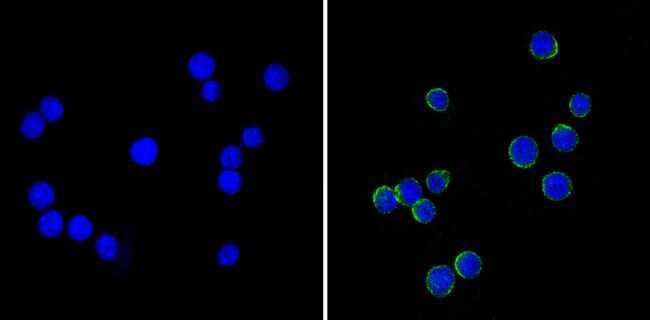
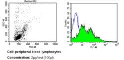
- Flow cytometry analysis of CD54 in PBMC cells (green) compared to an isotype control (blue). Human blood was collected, combined with a hydrophilic polysaccharide, centrifuged, transferred to a conical tube and washed with PBS. 50 µL of cell solution was added to each tube at a dilution of 2x10^7 cells/mL, followed by the addition of 50 µL of isotype control and primary antibody (Product # MA5-13021) at a dilution of 2 µg/test. Cells were incubated for 30 min at 4ºC and washed with a cell buffer, followed by incubation with a DyLight 488-conjugated secondary antibody for 30 min at 4ºC in the dark. FACS analysis was performed using 400 µL of cell buffer.

- Submitted by
- Invitrogen Antibodies (provider)
- Main image
- Experimental details
- Flow cytometry analysis of CD54 in PBMC cells (green) compared to an isotype control (blue). Human blood was collected, combined with a hydrophilic polysaccharide, centrifuged, transferred to a conical tube and washed with PBS. 50 µL of cell solution was added to each tube at a dilution of 2x10^7 cells/mL, followed by the addition of 50 µL of isotype control and primary antibody (Product # MA5-13021) at a dilution of 2 µg/test. Cells were incubated for 30 min at 4ºC and washed with a cell buffer, followed by incubation with a DyLight 488-conjugated secondary antibody for 30 min at 4ºC in the dark. FACS analysis was performed using 400 µL of cell buffer.

Supportive validation
- Submitted by
- Invitrogen Antibodies (provider)
- Main image

- Experimental details
- NULL