 Explore
 Validate
 Learn
Learn Western blot
Antibody data
- Antibody Data
- Antigen structure
- References [2]
- Comments [0]
- Validations
- Western blot [1]
- Flow cytometry [1]
Submit
Validation data
Reference
Comment
Report error
- Product number
- ABIN1882096 - Provider product page

- Provider
- antibodies-online
- Product name
- anti-Intercellular Adhesion Molecule 1 (ICAM1) (C-Term), (AA 488-514) antibody
- Antibody type
- Polyclonal
- Antigen
- This ICAM1 antibody is generated from rabbits immunized with a KLH conjugated synthetic peptide between 488-514 amino acids from the C-terminal region of human ICAM1.
- Description
- This antibody is prepared by Saturated Ammonium Sulfate (SAS) precipitation followed by dialysis against PBS. Purified Rabbit Polyclonal Antibody (Pab)
- Reactivity
- Human
- Host
- Rabbit
- Epitope
- C-Term,AA 488-514
- Antibody clone number
- RB20472
- Vial size
- 400 μL
- Concentration
- 2 mg/mL
Submitted references Myocardin-related transcription factor A mediates OxLDL-induced endothelial injury.
VLA molecule expression may be involved in the release of acute myeloid leukaemic cells from the bone marrow.
Fang F, Yang Y, Yuan Z, Gao Y, Zhou J, Chen Q, Xu Y
Circulation research 2011 Apr 1;108(7):797-807
Circulation research 2011 Apr 1;108(7):797-807
VLA molecule expression may be involved in the release of acute myeloid leukaemic cells from the bone marrow.
Denkers IA, de Jong-de Boer TJ, Beelen RH, Ossenkoppele GJ, Langenhuijsen MM
Leukemia research 1992;16(5):469-74
Leukemia research 1992;16(5):469-74
No comments: Submit comment
Supportive validation
- Submitted by
- antibodies-online (provider)
- Main image

- Experimental details
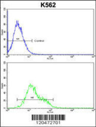
- ICAM1 Antibody (C-term) (ABIN1882096) flow cytometric analysis of k562 cells (bottom histogram) compared to a negative control cell (top histogram). FITC-conjugated goat-anti-rabbit secondary antibodies were used for the analysis.

Supportive validation
- Submitted by
- antibodies-online (provider)
- Main image
- Experimental details
- ICAM1 Antibody (C-term) (ABIN1882096) flow cytometric analysis of k562 cells (bottom histogram) compared to a negative control cell (top histogram). FITC-conjugated goat-anti-rabbit secondary antibodies were used for the analysis.
