 Explore
 Validate
 Learn
Learn Western blot
Antibody data
- Antibody Data
- Antigen structure
- References [1]
- Comments [0]
- Validations
- Western blot [3]
- Flow cytometry [1]
Submit
Validation data
Reference
Comment
Report error
- Product number
- 701254 - Provider product page

- Provider
- Invitrogen Antibodies
- Product name
- ICAM-1 Recombinant Rabbit Monoclonal Antibody (9H21L19)
- Antibody type
- Monoclonal
- Antigen
- Synthetic peptide
- Description
- This antibody is predicted to react with mouse, rat, non-human primate and rabbit based on sequence homology.
- Antibody clone number
- 9H21L19
- Concentration
- 0.5 mg/mL
Submitted references Proteomic analysis of exosomes derived from human lymphoma cells.
Yao Y, Wei W, Sun J, Chen L, Deng X, Ma L, Hao S
European journal of medical research 2015 Jan 29;20(1):8
European journal of medical research 2015 Jan 29;20(1):8
No comments: Submit comment
Supportive validation
- Submitted by
- Invitrogen Antibodies (provider)
- Main image

- Experimental details
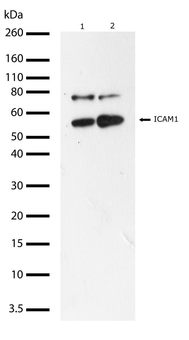
- Western blot analysis of ICAM-1 in whole cell extracts from Raji (lane 1), and K562 cells (lane 2) using an ICAM-1 recombinant rabbit monoclonal antibody (Product # 701254) at a dilution of 1 µg/mL. Samples were detected using chemiluminescence (ECL). Results show a band at ~58kDa.
- Submitted by
- Invitrogen Antibodies (provider)
- Main image

- Experimental details
- Western blot analysis was performed on whole cell extracts (30 µg lysate) of Raji (Lane 1), Mouse Spleen (Lane 2), Mouse Lung (lane 3), Rat Lung (lane 4) Mouse Kidney (lane 5) and Rat Kidney (lane 6). The blots were probed with Anti-ICAM-1 Recombinant Rabbit Monoclonal Antibody (Product # 701254, 1-2 µg/mL) and detected by chemiluminescence using Goat anti-Rabbit IgG (H+L) Superclonal™ Secondary Antibody, HRP conjugate (Product # A27036, 0.4 µg/mL, 1:2500 dilution). A 57 kDa band corresponding to ICAM-1 was observed across cell lines and tissues tested. Known quantity of protein samples were electrophoresed using Novex® NuPAGE® 12 % Bis-Tris gel (Product # NP0342BOX), XCell SureLock™ Electrophoresis System (Product # EI0002) and Novex® Sharp Pre-Stained Protein Standard (Product # LC5800). Resolved proteins were then transferred onto a nitrocellulose membrane with iBlot® 2 Dry Blotting System (Product # IB21001). The membrane was probed with the relevant primary and secondary Antibody following blocking with 5 % skimmed milk. Chemiluminescent detection was performed using Pierce™ ECL Western blotting Substrate (Product # 32106).

- Submitted by
- Invitrogen Antibodies (provider)
- Main image

- Experimental details
- Western blot was performed using Anti-ICAM-1 Recombinant Rabbit Monoclonal Antibody (9H21L19) (Product # 701254) and a 57-80 kDa band corresponding to Intercellular adhesion molecule 1 was observed across cell lines tested and upregulated in A549 with TNF-alpha treatment whereas downregulated in HT-1080 with PMA treatment.Whole cell extracts (30 µg lysate) of A549 (Lane 1), A549 treated with TNF-alpha (10 ng/mL for 6 hr) (Lane 2), HT-1080 (Lane 3), HT-1080 treated with PMA (50 nm for 4 hr) (Lane 4), MCF7 (Lane 5) and Raji (Lane 6) were electrophoresed using NuPAGE™ 4-12% Bis-Tris Protein Gel (Product # NP0322BOX). Resolved proteins were then transferred onto a nitrocellulose membrane (Product # IB23001) by iBlot® 2 Dry Blotting System (Product # IB21001). The blot was probed with the primary antibody (1 µg/mL) and detected by chemiluminescence with Goat anti-Rabbit IgG (H+L) Superclonal™ Recombinant Secondary Antibody, HRP (Product # A27036,1:20000 dilution) using the iBright FL 1000 (Product # A32752). Chemiluminescent detection was performed using SuperSignal™ West Pico PLUS Chemiluminescent Substrate (Product # 34580).

Supportive validation
- Submitted by
- Invitrogen Antibodies (provider)
- Main image

- Experimental details
- Flow cytometry analysis of ICAM-1 was done on Raji cells. Cells were fixed with 70% ethanol for 10 minutes, permeabilized with 0.25% Triton™ X-100 for 20 minutes, and blocked with 5% BSA for 30 minutes at room temperature. Cells were labeled with ABfinity™ ICAM-1 Recombinant Rabbit Monoclonal Antibody (701254, red histogram) or with rabbit isotype control (pink histogram) at 3-5 ug/million cells in 2.5% BSA. After incubation at room temperature for 2 hours, the cells were labeled with Alexa Fluor® 488 Goat Anti-Rabbit Secondary Antibody (A11008) at a dilution of 1:400 for 30 minutes at room temperature. The representative 10,000 cells were acquired and analyzed for each sample using an Attune® Acoustic Focusing Cytometer. The purple histogram represents unstained control cells and the green histogram represents no-primary-antibody control.
