Explore
 Validate
 Learn
Learn Other assay
Antibody data
- Antibody Data
- Antigen structure
- References [16]
- Comments [0]
- Validations
- Other assay [8]
Submit
Validation data
Reference
Comment
Report error
- Product number
- 14-0549-80 - Provider product page

- Provider
- Invitrogen Antibodies
- Product name
- CD54 (ICAM-1) Monoclonal Antibody (HA58), eBioscience™
- Antibody type
- Monoclonal
- Antigen
- Other
- Description
- Description: The HA58 monoclonal antibody reacts with human CD54 (InterCellular Adhesion Molecule-1, ICAM-1), a 90-110 kDa transmembrane glycoprotein expressed by monocytes, lymphocytes and endothelial cells. Expression of CD54 is upregulated on activated lymphocytes. Interaction of CD54 with its ligand CD11a is important in the inflammatory response.
- Antibody clone number
- HA58
- Concentration
- 0.5 mg/mL
Submitted references Pomalidomide restores immune recognition of primary effusion lymphoma through upregulation of ICAM-1 and B7-2.
Preservation of microvascular barrier function requires CD31 receptor-induced metabolic reprogramming.
CD54-NOTCH1 axis controls tumor initiation and cancer stem cell functions in human prostate cancer.
The critical role of SENP1-mediated GATA2 deSUMOylation in promoting endothelial activation in graft arteriosclerosis.
Type I IFNs induce anti-tumor polarization of tumor associated neutrophils in mice and human.
A Quantitative Perspective on Surface Marker Selection for the Isolation of Functional Tumor Cells.
Effect of nicotine and porphyromonas gingivalis lipopolysaccharide on endothelial cells in vitro.
The small GTPase Rap1b negatively regulates neutrophil chemotaxis and transcellular diapedesis by inhibiting Akt activation.
Minimal encounter time and separation determine ligand-receptor binding in cell adhesion.
Expression of endothelia and lymphocyte adhesion molecules in bronchus-associated lymphoid tissue (BALT) in adult human lung.
Tuning the formation and rupture of single ligand-receptor bonds by hyaluronan-induced repulsion.
Requirements for ICAM-1 immunogene therapy of lymphoma.
PR-39 and PR-11 peptides inhibit ischemia-reperfusion injury by blocking proteasome-mediated I kappa B alpha degradation.
PR-39 and PR-11 peptides inhibit ischemia-reperfusion injury by blocking proteasome-mediated I kappa B alpha degradation.
The human natural killer cell immune synapse.
The human natural killer cell immune synapse.
Shrestha P, Davis DA, Jaeger HK, Stream A, Aisabor AI, Yarchoan R
PLoS pathogens 2021 Jan;17(1):e1009091
PLoS pathogens 2021 Jan;17(1):e1009091
Preservation of microvascular barrier function requires CD31 receptor-induced metabolic reprogramming.
Cheung KCP, Fanti S, Mauro C, Wang G, Nair AS, Fu H, Angeletti S, Spoto S, Fogolari M, Romano F, Aksentijevic D, Liu W, Li B, Cheng L, Jiang L, Vuononvirta J, Poobalasingam TR, Smith DM, Ciccozzi M, Solito E, Marelli-Berg FM
Nature communications 2020 Jul 17;11(1):3595
Nature communications 2020 Jul 17;11(1):3595
CD54-NOTCH1 axis controls tumor initiation and cancer stem cell functions in human prostate cancer.
Li C, Liu S, Yan R, Han N, Wong KK, Li L
Theranostics 2017;7(1):67-80
Theranostics 2017;7(1):67-80
The critical role of SENP1-mediated GATA2 deSUMOylation in promoting endothelial activation in graft arteriosclerosis.
Qiu C, Wang Y, Zhao H, Qin L, Shi Y, Zhu X, Song L, Zhou X, Chen J, Zhou H, Zhang H, Tellides G, Min W, Yu L
Nature communications 2017 Jun 1;8:15426
Nature communications 2017 Jun 1;8:15426
Type I IFNs induce anti-tumor polarization of tumor associated neutrophils in mice and human.
Andzinski L, Kasnitz N, Stahnke S, Wu CF, Gereke M, von Köckritz-Blickwede M, Schilling B, Brandau S, Weiss S, Jablonska J
International journal of cancer 2016 Apr 15;138(8):1982-93
International journal of cancer 2016 Apr 15;138(8):1982-93
A Quantitative Perspective on Surface Marker Selection for the Isolation of Functional Tumor Cells.
Cahall CF, Lilly JL, Hirschowitz EA, Berron BJ
Breast cancer : basic and clinical research 2015;9(Suppl 1):1-11
Breast cancer : basic and clinical research 2015;9(Suppl 1):1-11
Effect of nicotine and porphyromonas gingivalis lipopolysaccharide on endothelial cells in vitro.
An N, Andrukhov O, Tang Y, Falkensammer F, Bantleon HP, Ouyang X, Rausch-Fan X
PloS one 2014;9(5):e96942
PloS one 2014;9(5):e96942
The small GTPase Rap1b negatively regulates neutrophil chemotaxis and transcellular diapedesis by inhibiting Akt activation.
Kumar S, Xu J, Kumar RS, Lakshmikanthan S, Kapur R, Kofron M, Chrzanowska-Wodnicka M, Filippi MD
The Journal of experimental medicine 2014 Aug 25;211(9):1741-58
The Journal of experimental medicine 2014 Aug 25;211(9):1741-58
Minimal encounter time and separation determine ligand-receptor binding in cell adhesion.
Robert P, Nicolas A, Aranda-Espinoza S, Bongrand P, Limozin L
Biophysical journal 2011 Jun 8;100(11):2642-51
Biophysical journal 2011 Jun 8;100(11):2642-51
Expression of endothelia and lymphocyte adhesion molecules in bronchus-associated lymphoid tissue (BALT) in adult human lung.
Kawamata N, Xu B, Nishijima H, Aoyama K, Kusumoto M, Takeuchi T, Tei C, Michie SA, Matsuyama T
Respiratory research 2009 Oct 22;10(1):97
Respiratory research 2009 Oct 22;10(1):97
Tuning the formation and rupture of single ligand-receptor bonds by hyaluronan-induced repulsion.
Robert P, Sengupta K, Puech PH, Bongrand P, Limozin L
Biophysical journal 2008 Oct;95(8):3999-4012
Biophysical journal 2008 Oct;95(8):3999-4012
Requirements for ICAM-1 immunogene therapy of lymphoma.
Kanwar JR, Berg RW, Yang Y, Kanwar RK, Ching LM, Sun X, Krissansen GW
Cancer gene therapy 2003 Jun;10(6):468-76
Cancer gene therapy 2003 Jun;10(6):468-76
PR-39 and PR-11 peptides inhibit ischemia-reperfusion injury by blocking proteasome-mediated I kappa B alpha degradation.
Bao J, Sato K, Li M, Gao Y, Abid R, Aird W, Simons M, Post MJ
American journal of physiology. Heart and circulatory physiology 2001 Dec;281(6):H2612-8
American journal of physiology. Heart and circulatory physiology 2001 Dec;281(6):H2612-8
The human natural killer cell immune synapse.
Davis DM, Chiu I, Fassett M, Cohen GB, Mandelboim O, Strominger JL
Proceedings of the National Academy of Sciences of the United States of America 1999 Dec 21;96(26):15062-7
Proceedings of the National Academy of Sciences of the United States of America 1999 Dec 21;96(26):15062-7
No comments: Submit comment
Supportive validation
- Submitted by
- Invitrogen Antibodies (provider)
- Main image

- Experimental details
- NULL
- Submitted by
- Invitrogen Antibodies (provider)
- Main image
- Experimental details
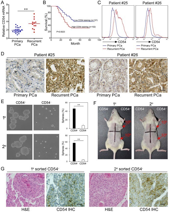
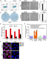
- Figure 3 Clinical Relevance of CD54 Expression. ( a ) Relative CD54 mRNA expression in primary (n=24) and recurrent tumors (n=12) from prostate cancer patients. ( b ) Relative CD54 mRNA expression correlated with mortality of patients with prostate cancer by Kaplan-Meier survival analysis. ( c ) CD54 expression in primary and recurrent tumors from two prostate cancer patients. ( d ) Immunohistochemical analysis of in situ CD54 expression in primary and recurrent tumors from two prostate cancer patients. ( e ) Representative light micrograph of the sphere-forming capacity of CD54 + and CD54 - sorted cells isolated from primary and secondary xenograft tumors of prostate cancer patients and quantification of the results. ( f ) Tumor-initiating capacity of differing numbers of different quadrants of CD54 + and CD54 - tumor cells isolated from primary and secondary prostate cancer patient xenograft tumors of NOD/SCID mice 6 weeks after injection. Red numbers indicate number of cells injected, and red arrows indicate tumor formation. ( g ) Immunohistochemical analysis of in situ CD54 expression in both primary and secondary tumor samples collected from xenografted CD54 + tumors. H&E stained tissues are shown as a reference (scale bar=50 mum). ** P < 0.01.
- Submitted by
- Invitrogen Antibodies (provider)
- Main image
- Experimental details
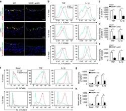
- Figure 3 Loss of endothelial SENP1 inhibits EC activation. ( a ) Grafts from WT or SENP1-ecKO mice were harvested 3 days post-transplantation. The induction of endothelial adhesion molecules was demonstrated by immunofluorescence staining of ICAM-1, VCAM-1, or P-selectin and PECAM-1 with DAPI labelling of the nuclei. Bar represents 50 mum. ( b - e ) Attenuated induction of adhesion molecules in SENP1-ecKO MAECs. Flow cytometry analysis of ICAM-1, VCAM-1 and P-selectin in MAECs isolated from WT or SENP1-ecKO mice after TNF or IL-1beta treatment. Representative histograms are shown in ( b ) with the quantification of mean intensity in ( c - e ). ( f - h ) Overexpression of the catalytically inactive form of SENP1 (SENP1-Mut) inhibits the induction of adhesion molecules in HUVECs. HUVECs were infected by Ad-SENP1-Mut or vector control (Ad-LacZ) for 24 h, treated with pro-inflammatory cytokines and analysed by flow cytometry in the same way as MAECs. Representative histograms of ICAM-1 and VCAM-1 are shown in ( f ) with the quantification of mean intensity in ( g , h ). Data are presented as the mean+-s.e.m. from at least three independent experiments. * P
- Submitted by
- Invitrogen Antibodies (provider)
- Main image

- Experimental details
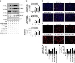
- Figure 8 GATA2 SUMOylation inhibits its DNA binding activity. ( a ) SUMO conjugation reduces GATA2 DNA binding activity. GATA2-WT, GATA2-2KR, SUMO-GATA2 or control constructs were transfected into 293 T cells, and nuclear extracts were processed with EMSA using a GATA2-specific oligonucleotide probe (top). Input of GATA2 was detected by western blotting with anti-Flag antibody (bottom). ( b ) Overexpression of catalytic inactive form of SENP1 (SENP1-Mut) inhibits recruitment of GATA2 to the promoter of ICAM-1, VCAM-1 and E-selectin. HUVECs were infected by Ad-SENP1-Mut or Ad-LacZ and then treated with TNF. Nuclear extracts were then subjected to ChIP assay with the anti-GATA2 antibody followed by quantitative real-time PCR for the promoter sequences of ICAM-1, VCAM-1 and E-selectin containing a GATA2 binding site. Quantitative results are shown as the ratio of ChIP to input values. Data are presented as the mean+-s.e.m. from three independent experiments. * P

- Submitted by
- Invitrogen Antibodies (provider)
- Main image
- Experimental details
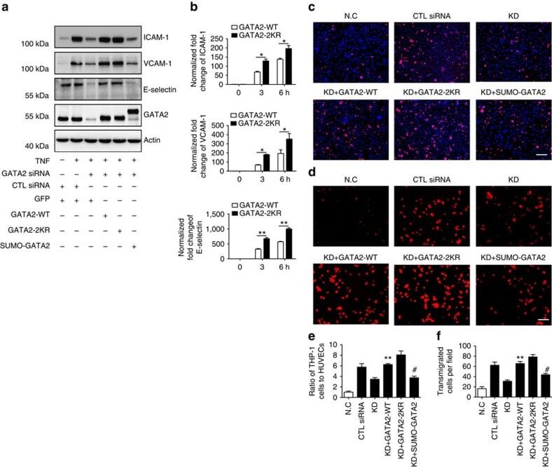
- Figure 9 The critical role of GATA2 SUMOylation in EC activation. ( a ) GATA2 SUMOylation regulates the expression of endothelial adhesion molecules. HUVECs were transfected by GATA2 siRNA or control siRNA for 48 h followed by infection with Ad-GATA2-WT, Ad-GATA2-KR, Ad-SUMO-GATA2 or their vector control (GFP) as indicated. HUVECs were treated with TNF or vehicle control for 24 h, and the cell lysates were then subjected to western blotting with anti-ICAM-1, anti-VCAM-1, anti-E-selectin or anti-GATA2 antibodies. Actin was used as a loading control. ( b ) GATA2 SUMOylation site mutation increases endothelial adhesion molecule induction by TNF at the early phase. The expression of endothelial adhesion molecules was determined by quantitative real-time PCR in HUVECs with GATA2-WT or GATA2-2KR reconstitution as described in ( a ). Data are presented as the mean+-s.e.m. from at least three independent experiments. * P
- Submitted by
- Invitrogen Antibodies (provider)
- Main image

- Experimental details
- Fig. 1 CD31 interactions promote the recovery of endothelial integrity following endothelial contraction induced by MHC molecule triggering. a - d Following MHC or ICAM-1 and/or CD31 antibody-mediated co-ligation for 30 min, EC were fixed and stained with rhodamine-phalloidin. Images taken on EC monolayers seeded at identical density are shown in ( a , b ). The average F-actin intensity per cell of three independent experiments is shown in ( c , d ). Scale bar, 20 mum. ( n = 3 biologically independent samples, N = 3 independent experiments, data are mean +- SD). One-way Anova with Tuckey post-hoc test. MHC vs all **** p < 0.0001, MHC + CD31 vs Isc **** p < 0.0001, MHC + CD31 vs all ****p < 0.0001. e Western blot (WB) analysis of Erk activation by WT and cd31 -/- EC 30 min after MHC stimulation. The bar graph shows relative protein expression +- SEM. N = 3 independent experiments (data are mean +- SD). One-way Anova with Tuckey post-hoc test. cd31 -/- MHC vs cd31 -/- IsC *** p = 0.0002, cd31 -/- MHC vs all **** p < 0.0001. f Western blot (WB) analysis of RhoA activation by WT and cd31 -/- EC 30 min after MHC stimulation. The bar graph shows relative protein expression +- SEM. N = 3 independent experiments (data are mean +- SD). One-way Anova with Tuckey post-hoc test. cd31 -/- 15' vs all *** p = 0.0003, cd31 -/- 30' vs all **** p < 0.0001. g Immunoprecipitation of CD31 molecules from WT EC exposed to MHC/ICAM-1 stimulation for 30 min followed by immunoblotting with an anti-pho

- Submitted by
- Invitrogen Antibodies (provider)
- Main image
- Experimental details
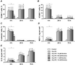
- Figure 5 Effect of nicotine on the P. gingivalis LPS-induced protein expression of pro-inflammatory mediators in HUVECs. HUVECs were stimulated by P. gingivalis LPS in the presence or absence of nicotine (10 uM-10 mM) for 4, 24, and 72 h. After stimulation, the surface expression levels of ICAM-1 (A), VCAM-1 (B), and E-selectin (C) were measured by flow cytometry, and the quantity of MCP-1 (D) and IL-8 (E) in conditioned media was measured by ELISA. Each value represents mean +-SD of three independent assays. Non-stimulated HUVECs were used as a control. The protein expression levels of pro-inflammatory mediators were not analyzed after stimulation with 10-mM nicotine for 24 and 72 h because the cells were not viable. * - significantly different between groups, p

- Submitted by
- Invitrogen Antibodies (provider)
- Main image
- Experimental details
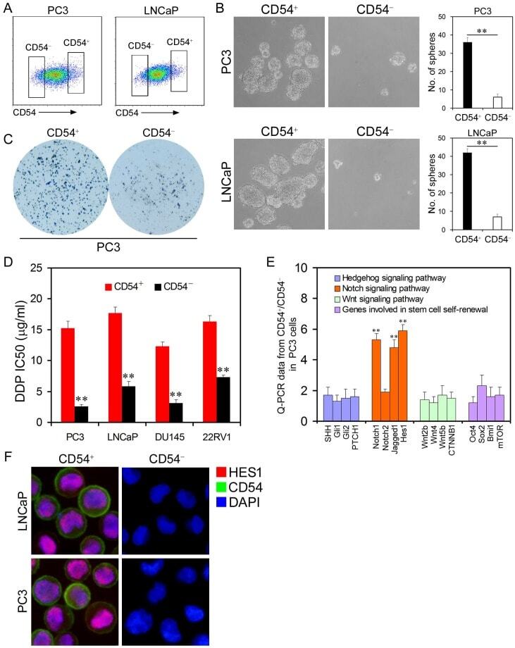
- Figure 2 Cancer Stem Cell Traits of Differential Populations of CD54 + and CD54 - Cells. ( a ) FACS histograms show separate gating for the isolation of CD54 + and CD54 - PC3 and LNCaP cells. ( b ) Representative light micrograph fields and comparative quantification of the sphere-forming capacity of CD54 + and CD54 - LNCaP and PC3 cells. ( c ) Representative microscope fields from colony formation assays for CD54 + and CD54 - PC3 cells. ( d ) The cisplatin (DDP) IC 50 of CD54 + and CD54 - cells from the PC3, LNCaP, DU145, and 22RV1 prostate cancer cell lines. ( e ) q-PCR analysis of the expression of Hedgehog, Notch, and Wnt signaling pathway genes as well as genes involved in stem cell self-renewal in CD54 + and CD54 - PC3 cells. ( f ) Representative micrographs of in situ protein expression of the key signaling pathway components HES1 and CD54 in DAPI-stained LNCaP and PC3 cells. ** P < 0.01.