 Explore
 Validate
 Learn
Learn Other assay
Antibody data
- Antibody Data
- Antigen structure
- References [3]
- Comments [0]
- Validations
- Other assay [5]
Submit
Validation data
Reference
Comment
Report error
- Product number
- BMS108 - Provider product page

- Provider
- Invitrogen Antibodies
- Product name
- CD54 (ICAM-1) Monoclonal Antibody (RR1/1), eBioscience™
- Antibody type
- Monoclonal
- Antigen
- Other
- Description
- Description: Recognizes the D1 domain of human CD54.
- Antibody clone number
- RR1/1
- Concentration
- 0.5 mg/mL
Submitted references Assessment of ICAM-1 N-glycoforms in mouse and human models of endothelial dysfunction.
Hydrogen peroxide regulates endothelial surface N-glycoforms to control inflammatory monocyte rolling and adhesion.
Tumor- and cytokine-primed human natural killer cells exhibit distinct phenotypic and transcriptional signatures.
Regal-McDonald K, Somarathna M, Lee T, Litovsky SH, Barnes J, Peretik JM, Traylor JG Jr, Orr AW, Patel RP
PloS one 2020;15(3):e0230358
PloS one 2020;15(3):e0230358
Hydrogen peroxide regulates endothelial surface N-glycoforms to control inflammatory monocyte rolling and adhesion.
McDonald KR, Hernandez-Nichols AL, Barnes JW, Patel RP
Redox biology 2020 Jul;34:101498
Redox biology 2020 Jul;34:101498
Tumor- and cytokine-primed human natural killer cells exhibit distinct phenotypic and transcriptional signatures.
Sabry M, Zubiak A, Hood SP, Simmonds P, Arellano-Ballestero H, Cournoyer E, Mashar M, Pockley AG, Lowdell MW
PloS one 2019;14(6):e0218674
PloS one 2019;14(6):e0218674
No comments: Submit comment
Supportive validation
- Submitted by
- Invitrogen Antibodies (provider)
- Main image
- Experimental details
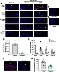
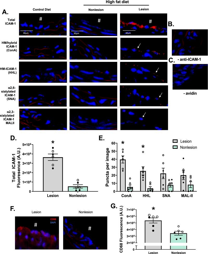
- Fig 2 HM epitopes co-localize with ICAM-1 in high fat-induced mouse atherosclerosis. Total, HM / hybrid, alpha-2,6-sialylated, and alpha-2,3-sialylated ICAM-1 were measured in the innominate and left carotid arteries from ApoE-/- mice fed a normal or high fat diet. A) Shown are representative images of innominate arteries from paired lesion and non-lesion areas of the same vessel section. Red staining represents total ICAM-1, red puncta represent positive PLA staining for specific ICAM-1 N-glycoforms (indicated by arrows), and blue staining represents DAPI. # indicates the lumen of each vessel. Panels B and C show PLA staining of lesion areas when the anti-ICAM-1 antibody or avidin were excluded. Panel D shows total ICAM-1 staining in lesion versus non-lesion areas. *p
- Submitted by
- Invitrogen Antibodies (provider)
- Main image
- Experimental details
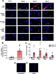
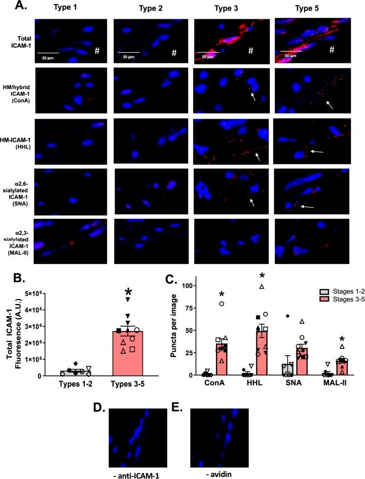
- Fig 5 HM / hybrid ICAM-1 is increased in human atherosclerosis. Panel A shows representative images of total ICAM-1 (red staining) and specified N-glycoforms in human vessels with lesions spanning types 1-5. Red puncta represent positive PLA staining (as indicated by arrows). Panel B shows the quantification of total ICAM-1 in early (1-2) and late (3-5) disease stages. Each symbol represents a different patient, with same symbol representing multiple vessels from the same patient. Data are mean +- SEM, n = 7-10. * p
- Submitted by
- Invitrogen Antibodies (provider)
- Main image
- Experimental details
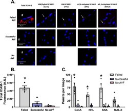
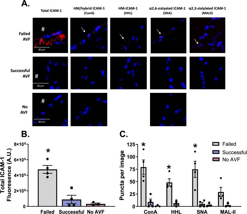
- Fig 7 HM / hybrid, HM, alpha-2,6-sialylated, ICAM-1 are increased CKD patients with failed arteriovenous fistulas. Panel A shows total ICAM-1 (red staining) and specified N-glycoforms in vessels from CKD patients with failed or successful AVF creation. Red puncta represent positive PLA staining (as indicated by arrows). B) Quantification of total ICAM-1 signal in failed and successful AVF samples (n = 4 each). Error bars are mean +- SEM; * p
- Submitted by
- Invitrogen Antibodies (provider)
- Main image
- Experimental details
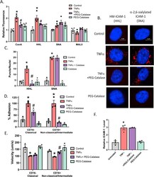
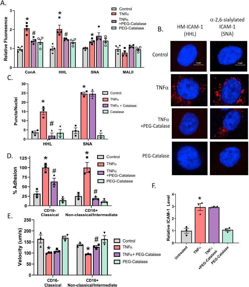
- Fig. 4 TNFalpha-induced HM-ICAM-1 formation and alpha-mannosidase activity inhibition can be reversed with PEG-Catalase. A. HUVECs treated with 100 units of PEG-catalase 30 min prior to TNFalpha treatment and lectin staining with ConA, HHL, SNA, and MAL-II was performed. * = p
- Submitted by
- Invitrogen Antibodies (provider)
- Main image
- Experimental details
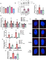
- Fig. 5 ERO1-alpha inhibition abrogates TNFalpha-inhibition of alpha mannosidase activity and HM N-glycan formation. A. HUVECs were pretreated with either 10 muM GKT137831 or 5 muM EN460 to inhibit NOX4, ERO1-alpha, respectively, prior to TNFalpha treatment and alpha-mannosidase activity measured. * = p
