Explore
 Validate
 Learn
Learn Flow cytometry
Antibody data
- Antibody Data
- Antigen structure
- References [1]
- Comments [0]
- Validations
- Flow cytometry [2]
- Other assay [1]
Submit
Validation data
Reference
Comment
Report error
- Product number
- MA5-28553 - Provider product page

- Provider
- Invitrogen Antibodies
- Product name
- CD54 (ICAM-1) Monoclonal Antibody (1H4), Alexa Fluor™ 700
- Antibody type
- Monoclonal
- Antigen
- Other
- Description
- This antibody recognizes an extracellular epitope of CD54 (ICAM-1), a 85-110 kDa type I transmembrane glycoprotein (receptor for rhinovirus) expressed on activated endothelial cells, T lymphocytes, B lymphocytes, monocytes, macrophages, granulocytes and dendritic cells; the expression of CD54 is upregulated by activation. Recommended dilution is for 100 µL of whole blood or 1x10^6 cells in suspension.
- Reactivity
- Human
- Host
- Mouse
- Conjugate
- Near infrared dye
- Isotype
- IgG
- Antibody clone number
- 1H4
- Vial size
- 100 Tests
- Storage
- 4°C, store in dark, DO NOT FREEZE!
Submitted references Colonization of dermal arterioles by Neisseria meningitidis provides a safe haven from neutrophils.
Manriquez V, Nivoit P, Urbina T, Echenique-Rivera H, Melican K, Fernandez-Gerlinger MP, Flamant P, Schmitt T, Bruneval P, Obino D, Duménil G
Nature communications 2021 Jul 27;12(1):4547
Nature communications 2021 Jul 27;12(1):4547
No comments: Submit comment
Supportive validation
- Submitted by
- Invitrogen Antibodies (provider)
- Main image

- Experimental details
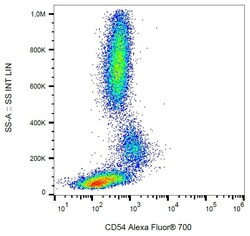
- Flow cytometry (surface staining) of human peripheral blood with anti-CD54 (1H4) Alexa Fluor® 700 Monoclonal antibody (Product # MA5-28553).
- Conjugate
- Near infrared dye

- Submitted by
- Invitrogen Antibodies (provider)
- Main image
- Experimental details
- Flow cytometry (surface staining) of human peripheral blood with anti-CD54 (1H4) Alexa Fluor® 700 Monoclonal antibody (Product # MA5-28553).

Supportive validation
- Submitted by
- Invitrogen Antibodies (provider)
- Main image
- Experimental details
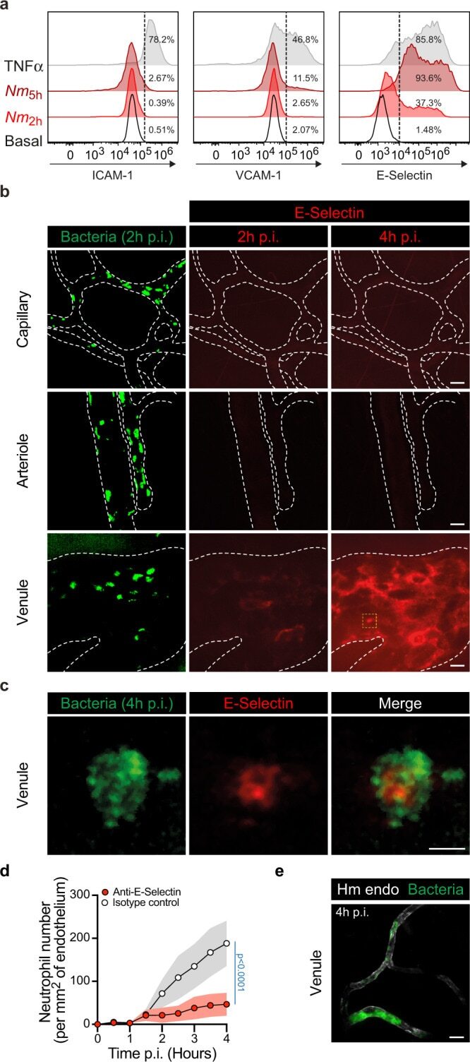
- Fig. 7 E-selectin endothelium surface expression is differentially upregulated according to the vascular bed upon infection. a Flow cytometry analysis of cell surface expression of ICAM-1, VCAM-1, and E-selectin (CD62E) on HUVEC cells under resting conditions (Basal, open histograms) or following infection with Neisseria meningitidis for 2 h ( Nm 2h , red histograms) or 5 h ( Nm 5h , dark-red histograms) or an overnight incubation with 20 ng ml -1 TNFalpha gray histograms). Data are representatives of N = 3 independent experiments. The percentages of positive cells (above the dashed lines) are shown per condition and marker. b In vivo expression of E-selectin at the surface of different human vessel types (capillary, arteriole, and venule) following two (2 h p.i.) and four (4 h p.i.) hours of infection. Images (maximum intensity z-projection) are representative of N = 3 infected mice imaged independently. GFP-expressing Neisseria meningitidis appears in green and E-selectin in red following in vivo labeling by i.v. injection of PE-labeled anti-CD62E monoclonal antibody. Dashed lines delineate human vessels (UEA-1 lectin). Scale bar, 20 um. c High-magnification view (yellow dashed square in panel b ) of a bacterial microcolony at 4 h p.i. on the venular endothelium surface and the local upregulation of E-selectin expression. Scale bar, 5 um. d Movies obtained from intravital imaging were used to quantify the numbers of neutrophils per square millimeter of venular endothelium d
- Conjugate
- Near infrared dye