Explore
 Validate
 Learn
Learn Western blot
 Immunohistochemistry
Antibody data
- Antibody Data
- Antigen structure
- References [5]
- Comments [0]
- Validations
- Immunohistochemistry [1]
Submit
Validation data
Reference
Comment
Report error
- Product number
- HPA004877 - Provider product page

- Provider
- Atlas Antibodies
- Proper citation
- Atlas Antibodies Cat#HPA004877, RRID:AB_1846317
- Product name
- Anti-ICAM1
- Antibody type
- Polyclonal
- Description
- Polyclonal Antibody against Human ICAM1, Gene description: intercellular adhesion molecule 1, Alternative Gene Names: BB2, CD54, Validated applications: IHC, WB, Uniprot ID: P05362, Storage: Store at +4°C for short term storage. Long time storage is recommended at -20°C.
- Reactivity
- Human
- Host
- Rabbit
- Conjugate
- Unconjugated
- Isotype
- IgG
- Vial size
- 100 µl
- Concentration
- 0.2 mg/ml
- Storage
- Store at +4°C for short term storage. Long time storage is recommended at -20°C.
- Handling
- The antibody solution should be gently mixed before use.
Submitted references ICAM1 antibody drug conjugates exert potent antitumor activity in papillary and anaplastic thyroid carcinoma
Dynamic Glycoprotein Hyposialylation Promotes Chemotherapy Evasion and Metastatic Seeding of Quiescent Circulating Tumor Cell Clusters in Breast Cancer
ICAM1 initiates CTC cluster formation and trans-endothelial migration in lung metastasis of breast cancer
Persistence of endothelial thrombomodulin in a patient with infectious purpura fulminans treated with protein C concentrate
Variance decomposition of protein profiles from antibody arrays using a longitudinal twin model
Zhang P, Tao C, Shimura T, Huang A, Kong N, Dai Y, Yao S, Xi Y, Wang X, Fang J, Moses M, Guo P
iScience 2023;26(8):107272
iScience 2023;26(8):107272
Dynamic Glycoprotein Hyposialylation Promotes Chemotherapy Evasion and Metastatic Seeding of Quiescent Circulating Tumor Cell Clusters in Breast Cancer
Dashzeveg N, Jia Y, Zhang Y, Gerratana L, Patel P, Shajahan A, Dandar T, Ramos E, Almubarak H, Adorno-Cruz V, Taftaf R, Schuster E, Scholten D, Sokolowski M, Reduzzi C, El-Shennawy L, Hoffmann A, Manai M, Zhang Q, D'Amico P, Azadi P, Colley K, Platanias L, Shah A, Gradishar W, Cristofanilli M, Muller W, Cobb B, Liu H
Cancer Discovery 2023;13(9):2050-2071
Cancer Discovery 2023;13(9):2050-2071
ICAM1 initiates CTC cluster formation and trans-endothelial migration in lung metastasis of breast cancer
Taftaf R, Liu X, Singh S, Jia Y, Dashzeveg N, Hoffmann A, El-Shennawy L, Ramos E, Adorno-Cruz V, Schuster E, Scholten D, Patel D, Zhang Y, Davis A, Reduzzi C, Cao Y, D’Amico P, Shen Y, Cristofanilli M, Muller W, Varadan V, Liu H
Nature Communications 2021;12(1)
Nature Communications 2021;12(1)
Persistence of endothelial thrombomodulin in a patient with infectious purpura fulminans treated with protein C concentrate
Bendapudi P, Robbins A, LeBoeuf N, Pozdnyakova O, Bhatt A, Duke F, Sells R, McQuiston J, Humrighouse B, Rouaisnel B, Colling M, Stephenson K, Saavedra A, Losman J
Blood Advances 2018;2(21):2917-2921
Blood Advances 2018;2(21):2917-2921
Variance decomposition of protein profiles from antibody arrays using a longitudinal twin model
Kato B, Nicholson G, Neiman M, Rantalainen M, Holmes C, Barrett A, Uhlén M, Nilsson P, Spector T, Schwenk J
Proteome Science 2011;9(1):73
Proteome Science 2011;9(1):73
No comments: Submit comment
Supportive validation
- Submitted by
- Atlas Antibodies (provider)
- Enhanced method
- Orthogonal validation
- Main image

- Experimental details
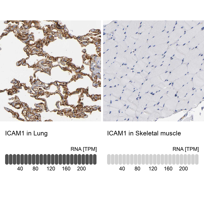
- Immunohistochemistry analysis in human lung and skeletal muscle tissues using HPA004877 antibody. Corresponding ICAM1 RNA-seq data are presented for the same tissues.
- Sample type
- Human
- Protocol
- Protocol